

doi: 10.1038/nprot.2006.229.
Determination of the folding of proteins as a function of denaturants, osmolytes or ligands using circular dichroism
Affiliations
- PMID: 17406529
- PMCID: PMC2728349
- DOI: 10.1038/nprot.2006.229
Item in Clipboard
Determination of the folding of proteins as a function of denaturants, osmolytes or ligands using circular dichroism
Nat Protoc.
2006.
Abstract
Circular dichroism (CD) is an excellent tool for examining the interactions and stability of proteins. This protocol covers methods to obtain and analyze circular dichroism spectra to measure changes in the folding of proteins as a function of denaturants, osmolytes or ligands. Applications include determination of the free energy of folding of a protein, the effects of mutations on protein stability and the estimation of binding constants for the interactions of proteins with other proteins, DNA or ligands, such as substrates or inhibitors. The experiments require 2-5 h.
Figures
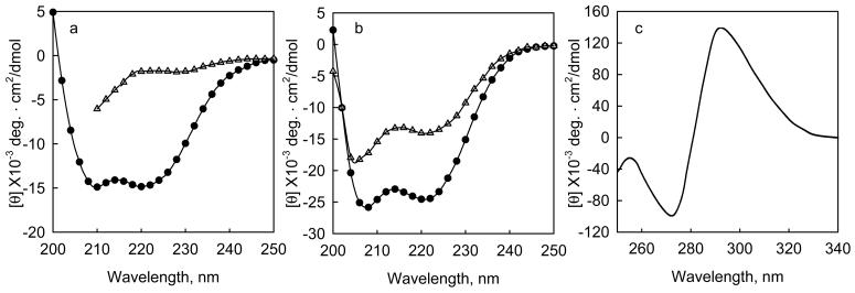
a, The change in mean residue ellipticity when Gn-HCl is added to a fragment of tropomodulin 1, Tmod1160-359, 0.24 mg/ml (10 μM) in 100 mM NaCl, 10 mM sodium phosphate, pH 6.5 at 10 °C. (o) native protein, (Δ) protein + 3.6 M Gn-HCl. b. The change in mean residue ellipticity when calcium binds to the calcium binding subunit of cardiac troponin, TnC, 0.2 mg/ml, at 2 °C in 50 mM NaC1, 2 mM HEPES, pH 7.0, 0.5 mM DTT, with (Δ) 2mM EDTA, or (•) 3 mM CaCl2. c, Induced molar ellipticity when saturating sodium folate binds to dihydrofolate reductase from e coli.. The enzyme is 18 μM in 100 mM NaCl, pH 7.2 at 27 °C.
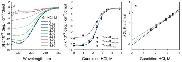
a, The mean residue ellipticity of three fragments of tropomodulin 1 as a function of Gn-HCl in 100 mM NaCl, 10 mM sodium phosphate, pH 6.5 at 10 °C. a, (black circles), Tmod1160-359, 0.24 mg/ml; (magenta triangles), residues Tmod1160-344, 0.17 mg/ml; (cyan squares), Tmod11-322, 0.22 mg/ml. The data were fit to equations 9-12 by non-linear least squares analysis. b, The data in panel a were transformed using equations 2, 3 and 5 to give a linear plot of ΔG as a function of denaturant. The free energy of folding equals the value of the y intercept.
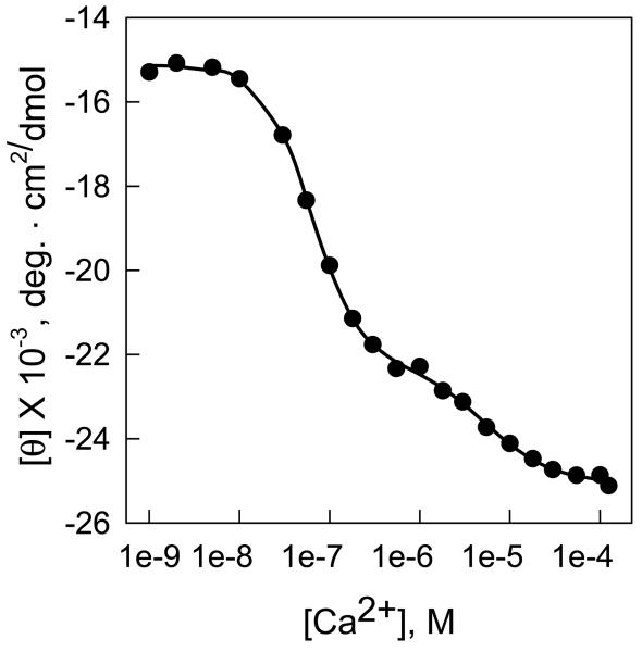
0.4 ml of chicken muscle Troponin C (TnC), 0.1 mg/ml, in 100 mM KCl, 50 mM Tris-HCl, pH 7.5, 0.9 mM EGTA, 0.9 mM nitriloacetic acid, pH 7.5 at 25 °C, in a 0.2 ml cuvette was titrated with Ca2+.
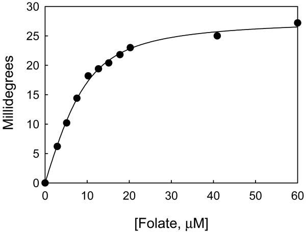
When folate binds to Dihydrofolate reductase from E. coli its absorption bands become optically active. (•) The change of the difference in ellipticity between 292.5and 270.0 nm of dihydrofolate reductase from E. coli as a function of increasing folate concentration. (line) The data fit by equation (23) to determine the number of binding sites/mole (0.95), the maximal ellipticity change, 28 millidegrees, and the dissociation constant, 3.1 μM. The enzyme was 9.2 μM in 0.1 N NaCl, pH 7.2, at 27 °C.
References
-
- Fowler VM, Greenfield NJ, Moyer J. Tropomodulin contains two actin filament pointed end-capping domains. J. Biol. Chem. 2003;278:40000–9. - PubMed
-
- Smith L, Greenfield NJ, Hitchcock-DeGregori SE. The effects of deletion of the amino-terminal helix on troponin C function and stability. J. Biol. Chem. 1994;269:9857–63. - PubMed
-
- Greenfield NJ, Williams MN, Poe M, Hoogsteen K. Circular dichroism studies of dihydrofolate reductase from a methotrexate-resistant strain of Escherichia coli. Biochemistry. 1972;11:4706–11. - PubMed
-
- Schellman JA. Macromolecular Binding. Biopolymers. 1975;14:999–1018.
Publication types
MeSH terms
Substances
Grants and funding
LinkOut - more resources
Full Text Sources
