. 2007 May;37(5):1334-42.
doi: 10.1002/eji.200636362.

A role for plasmacytoid dendritic cells in the rapid IL-18-dependent activation of NK cells following HSV-1 infection

Affiliations

A role for plasmacytoid dendritic cells in the rapid IL-18-dependent activation of NK cells following HSV-1 infection

Daniel P Barr et al. Eur J Immunol. 2007 May.

Abstract

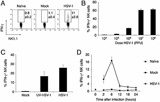
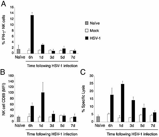
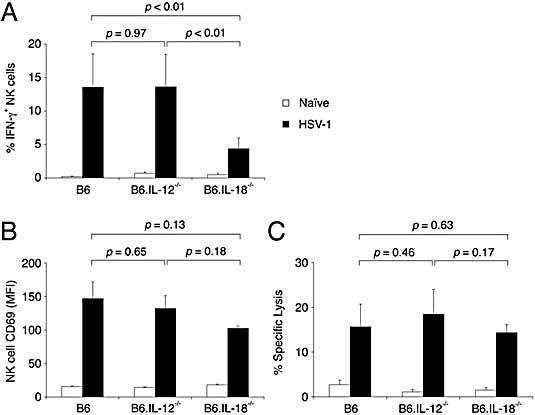
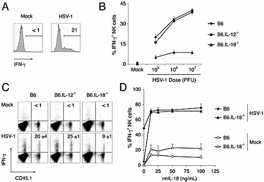
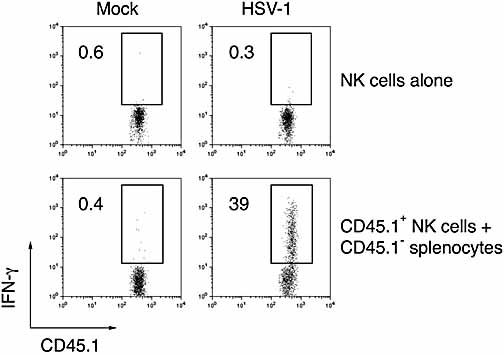
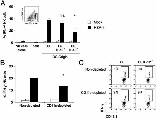
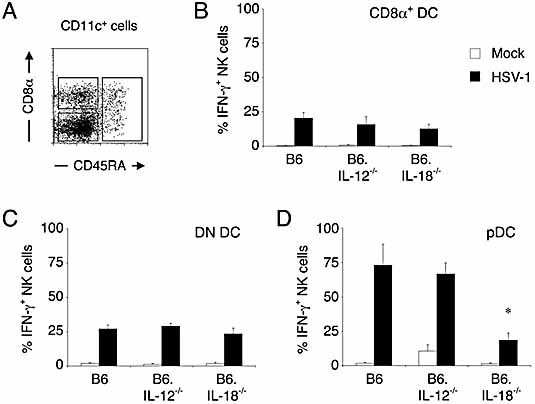
Natural killer (NK) cells play a crucial role in the initial response to viral infections but the mechanisms controlling their activation are unclear. We show a rapid and transient activation of NK cells that results in the production of IFN-gamma immediately following infection with herpes simplex virus type 1 (HSV-1). Activation of NK cells leading to synthesis of IFN-gamma was not mediated by a direct interaction with virus but required the presence of additional cell types and was largely dependent on the cytokine IL-18, but not IL-12. HSV-1-induced IFN-gamma expression by NK cells in vitro was impaired in spleen cultures depleted of CD11c(+) cells. Conversely, coculture of NK cells with virus-exposed conventional DC or plasmacytoid (p)DC restored the production of IFN-gamma, indicating that multiple DC subsets could mediate NK cell activation. While conventional DC populations stimulated NK cells independently of IL-18, they were less effective than pDC in promoting NK cell IFN-gamma expression. In contrast, the potent stimulation of NK cells by pDC was dependent on IL-18 as pDC from IL-18-deficient mice only activated a similar proportion of NK cells as conventional DC. These data identify IL-18 as a crucial factor for pDC-mediated NK cell regulation.


Figures

Figure 1
Figure 1
Immediate activation of NK cells following intravenous infection with HSV-1. Spleens were obtained from naïe B6 mice or 6 h after B6 mice were injected intravenously with (A) Vero cell lysate (Mock) or 106 PFU of HSV-1, (B) increasing doses of HSV-1, (C) Vero cell lysate (Mock), 106 PFU of HSV-1 or an equivalent amount of UV-inactivated HSV-1, and the proportion of NK cells producing IFN-γ determined by flow cytometry. (D) Spleens were obtained from naïe, mock-infected (Vero cell lysate) or HSV-1-infected (106 PFU) mice at the indicated times post-infection and the proportion of NK cells producing IFN-γ determined by flow cytometry. Data in panel (A) show representative dot plots of IFN-γ production by NK cells, and have been gated on CD3ε NK1.1+ lymphocytes. Data in panels (B) and (C) show the mean proportion of NK cells producing IFN-γ (± SD) and are representative of three independent experiments. Data in panel (D) show the mean proportion of NK cells producing IFN-γ (± SE) from three independent experiments
Figure 2
Figure 2
The NK cell response to HSV-1 is characterized by IFN-γ production, CD69 expression and increased YAC-1 cytolysis. Splenocytes were prepared from naïe (shaded column), mock-infected (unfilled columns) and HSV-1-infected (106 PFU, filled columns) mice at 6 h and 1, 3, 5 and 7 days post-infection. Cells were stained with anti-NK1.1-FITC and anti-CD3ε-APC and the expression of (A) intracellular IFN-γ and (B) cell surface CD69 by NK cells was assessed by flow cytometry. (C) Spleen cells were incubated with 51Cr-labeled targets at an effector to target ratio of 100:1 and specific lysis was determined. Columns represent mean ± SD of three mice at each time point. Data are representative of two independent experiments.
Figure 3
Figure 3
Rapid synthesis of IFN-γ by NK cells in response to HSV-1 infection is dependent on IL-18 in vivo. Splenocytes were prepared from B6, B6.IL-12–/– and B6.IL-18–/– mice that were naïe (unfilled columns) or infected (A) 6 h, or (B, C) 24 h earlier with 106 PFU of HSV-1 (filled columns). (A) Cells were incubated for 4 h in Brefeldin A and then stained with mAb specific for NK1.1, CD3ε and IFN-γ. The proportion of IFN-γ+ NK cells was subsequently determined by flow cytometry. Columns represent mean ± SD of five mice. Data are representative of at least three independent experiments. (B) Cells were stained with mAb specific for NK1.1, CD3ε and CD69. The expression of CD69 by NK cells was subsequently determined by flow cytometry. Columns represent mean ± SD of four mice. Data are representative of three independent experiments. (C) Spleen cells were incubated with 51Cr-labeled targets at an effector to target ratio of 100:1 and specific lysis determined. Columns represent mean ± SE of eight mice from two independent experiments. Statistical significance comparing results from infected animals is indicated.
Figure 4
Figure 4
Crucial role of IL-18 for HSV-1-induced IFN-γ production by NK cells. (A) Splenocytes from naïe mice were cultured for 8 h with Vero cell lysate (Mock) or 106 PFU of HSV-1. Cells were then cultured in Brefeldin A for 4 h, stained and the proportion of IFN-γ+ NK cells determined by flow cytometry. Representative histograms showing IFN-γ production after gating on CD3ε NK1.1+ cells. (B) The proportion of IFN-γ+ NK cells in cultures of B6 (diamonds), B6.IL-12–/– (squares) and B6.IL-18–/– (triangles) splenocytes after incubation with increasing doses of HSV-1. (C) Purified B6.CD45.1 NK cells (5 × 10⁴) were cocultured with 106 CD45.1 B6, B6.IL-12–/– or B6.IL-18–/– splenocytes with Mock inoculum or HSV-1. Values represent the proportion of CD45.1+ IFN-γ+ events. (D) IFN-γ production by NK cells in B6 (diamonds) and B6.IL-18–/– (triangles) splenocytes induced by Mock inoculum (unfilled) or HSV-1 (filled) in the presence of the indicated concentrations of recombinant mouse (rm)IL-18. (B–D) Data show mean ± SD from triplicate cultures. All data are representative of at least three independent experiments.
Figure 5
Figure 5
HSV-1-induced activation of NK cells in vitro requires other cell types. Purified CD45.1+ NK cells (5 × 10⁴) were cultured for 8 h with Vero cell lysate (Mock) or with 106 PFU HSV-1 in the presence or absence of 106 B6 splenocytes (CD45.1). After incubation in Brefeldin A for an additional 4 h, cells were labelled with anti-CD45.1 to identify purified NK cells and then assessed for IFN-γ production. Values represent the proportion of CD45.1+ IFN-γ+ events and are representative of at least three experiments.
Figure 6
Figure 6
DC-derived IL-18 regulates HSV-1-induced NK cell activation. (A) Splenic DC were purified from B6, B6.IL-12–/– and B6.IL-18–/– mice by cell sorting (insert) and incubated with purified NK cells (1 × 10⁴) with 106 PFU HSV-1 (filled columns) or Vero cell lysate (unfilled columns). Data are shown as mean ± SE. Statistical significance comparing results B6 DC to cytokine-deficient DC is indicated (n.s., not significant; *p 0.05). (B) Splenocytes from B6 mice or splenocytes depleted of CD11c-positive cells were cultured in the presence or absence of HSV-1 and the proportion of NK cells producing IFN-γ determined (mean ± SD) Statistical significance is indicated (*p <0.05). (C) Purified B6.CD45.1 NK cells were cocultured with CD11c-depleted and non-depleted splenocytes from B6 and B6.IL-12–/– mice in the presence of HSV-1 and then stained with anti-CD45.1 and anti-IFN-γ. Values represent the proportion of IFN-γ+ CD45.1+ NK cells determined by flow cytometry.
Figure 7
Figure 7
HSV-1-induced NK cell activation is mediated by multiple DC subsets. DC from naïe B6, B6.IL-12–/– and B6.IL-18–/– mice were enriched from splenocytes and stained with mAb specific for CD11c, CD8α and CD45RA. (A) Representative dot plot of CD11c+ cells from a B6 animal analysed for expression of CD8α and CD45RA. DC subsets were purified by sorting of CD11c+ splenocytes to obtain CD8α CD45RA (DN DC), CD8α+ CD45RA (CD8α+ DC) or CD45RA+ (pDC). Purified NK cells (1 × 10⁴) were incubated with (5 × 10⁴) purified (B) CD8α+ DC, (C) DN DC, or (D) pDC subsets in the presence of 106 PFU HSV-1 (filled columns) or Vero cell lysate (unfilled columns) for 6 to 8 h. Following incubation in Brefeldin A for an additional 4 h, cells were stained with anti-NK1.1 and anti-TCRβ. Cells were then fixed, permeabilised and stained for intracellular IFN-γ, and the proportion of NK cells that expressed IFN-γ determined by flow cytometry. The proportion of IFN-γ+ NK cells are expressed as mean ± SE from three independent experiments. Statistical significance comparing results from cultures of NK cells mixed with purified cytokine-deficient DC subsets to B6 DC subsets is indicated; *p <0.05.

References

    1. Yokoyama WM, Kim S, French AR. The dynamic life of natural killer cells. Annu. Rev. Immunol. 2004;22:405–429. - PubMed
    1. Karlhofer FM, Ribaudo RK, Yokoyama WM. MHC class I alloantigen specificity of Ly-49+ IL-2-activated natural killer cells. Nature. 1992;358:66–70. - PubMed
    1. Wagtmann N, Biassoni R, Cantoni C, Verdiani S, Malnati MS, Vitale M, Bottino C, et al. Molecular clones of the p58 NK cell receptor reveal immunoglobulin-related molecules with diversity in both the extra- and intracellular domains. Immunity. 1995;2:439–449. - PubMed
    1. Biron CA, Nguyen KB, Pien GC, Cousens LP, Salazar-Mather TP. Natural killer cells in antiviral defense: function and regulation by innate cytokines. Annu. Rev. Immunol. 1999;17:189–220. - PubMed
    1. Kobayashi M, Fitz L, Ryan M, Hewick RM, Clark SC, Chan S, Loudon R, et al. Identification and purification of natural killer cell stimulatory factor (NKSF), a cytokine with multiple biologic effects on human lymphocytes. J. Exp. Med. 1989;170:827–845. - PMC - PubMed

Publication types