. 2007 Jul 15;405(2):299-306.
doi: 10.1042/BJ20070051.

Enzymatic cleavage specificity of the proalpha1(V) chain processing analysed by site-directed mutagenesis

Affiliations

Enzymatic cleavage specificity of the proalpha1(V) chain processing analysed by site-directed mutagenesis

Christelle Bonod-Bidaud et al. Biochem J. .

Abstract

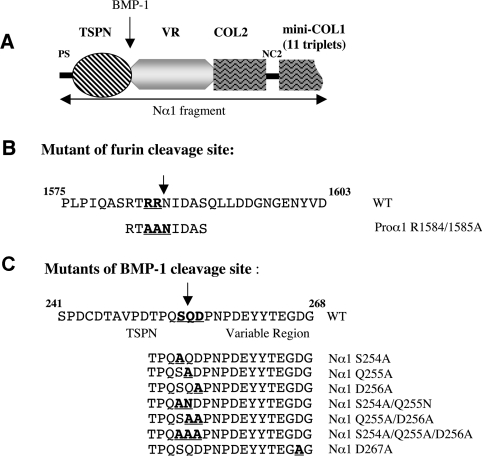
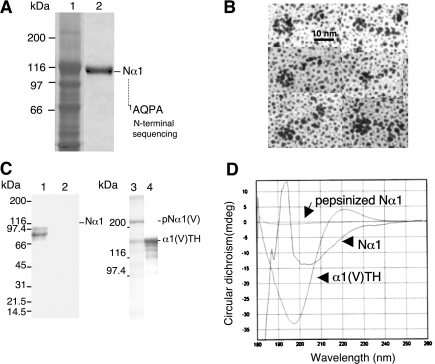
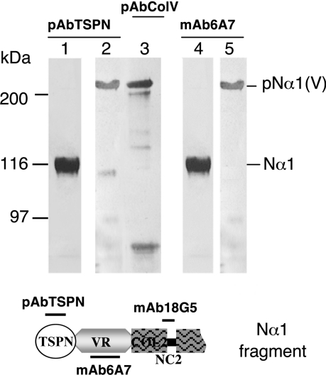
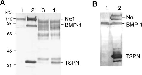
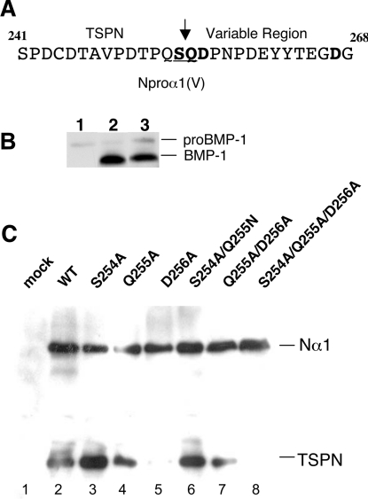
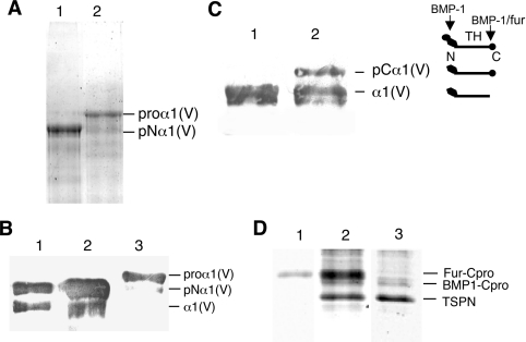
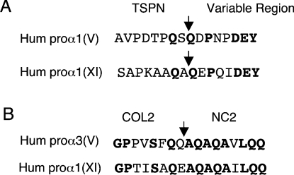
The proteolytic processing of procollagen V is complex and depends on the activity of several enzymes among which the BMP-1 (bone morphogenetic protein-1)/tolloid metalloproteinase and the furin-like proprotein convertases. Few of these processing interactions could have been predicted by analysing the presence of conserved consensus sequences in the proalpha1(V) chain. In the present study we opted for a cell approach that allows a straightforward identification of processing interactions. A construct encompassing the complete N-terminal end of the proalpha1(V) chain, referred to as Nalpha1, was recombinantly expressed to be used for enzymatic assays and for antibody production. Structural analysis showed that Nalpha1 is a monomer composed of a compact globule and an extended tail, which correspond respectively to the non-collagenous Nalpha1 subdomains, TSPN-1 (thrombospondin-1 N-terminal domain-like) and the variable region. Nalpha1 was efficiently cleaved by BMP-1 indicating that the triple helix is not required for enzyme activity. By mutating residues flanking the cleavage site, we showed that the aspartate residue at position P2' is essential for BMP-1 activity. BMP-1 activity at the C-terminal end of the procollagen V was assessed by generating a furin double mutant (R1584A/R1585A). We showed that, in absence of furin activity, BMP-1 is capable of processing the C-propeptide even though less efficiently than furin. Altogether, our results provide new relevant information on this complex and poorly understood mechanism of enzymatic processing in procollagen V function.


Figures

Figure 1
Figure 1. Constructs and mutants of the human proα1(V) chain
(A) Schematic representation of the N-propeptide construct referred to as Nα1. It consists of NC3, COL2, NC2 and 11 triplets from COL1. (B) Sequences of the C-propeptide encompassing the furin cleavage site and of the R1584A/R1585A mutant. The two arginine residues at positions 1584 and 1585 were mutated to alanine. (C) Sequences of the N-propeptide encompassing the BMP-1 cleavage site and of the BMP-1 cleavage site mutants. COL, collagenous domain; NC, non-collagenous domain. Mutated residues are underlined. Arrows indicate cleavage sites.
Figure 2
Figure 2. Production and characterization of Nα1 fragment and antibodies
(A) SDS/6% PAGE analysis of recombinant Nα1 fragment produced in HEK-293 cells. Electrophoretic patterns of serum-free medium from transfected cells (lane 1) and of the purified Nα1 fragment (lane 2). (B) Rotary-shadowing images of recombinant purified Nα1 fragment revealing the presence of a compact globular domain (TSPN-1 domain) and an extended rod-like domain (variable region). (C) SDS/PAGE (5–20% gradient) analysis of Nα1 (lane 1) and Nα1 digested with pepsin (lane 2) and SDS/6% PAGE analysis of α1(V) homotrimeric molecule treated without (lane 3) and with pepsin (lane 4). (D) CD spectra of α1(V)TH, Nα1 and Nα1 after pepsin digestion. Buffer baselines were systematically subtracted.
Figure 3
Figure 3. Western-blot characterization of the polyclonal antibodies to the TSPN-1 domain (pAbTSPN) and the mAb (mAb6A7) directed against the variable region of the Nα1 fragment
pAbTSPN (lanes 1 and 2) and the mAb 6A7 (lanes 4 and 5) recognized the complete proα1(V) chain (lanes 2 and 5) and the Nα1 fragment (lanes 1 and 4). Lane 3 shows proα1(V) chain recognition by polyclonal antibodies directed against the collagen V triple helix (from Novotec). Epitopes of the different antibodies are indicated on the schematic representation of the Nα1 fragment presented below. Left: running positions of protein standards are indicated in kDa.
Figure 4
Figure 4. BMP-1 cleavage of the N-propeptide domain of the proα1(V) chain
(A) Western-blot analysis of BMP-1 processing of the N-propeptide of the proα1(V) chain in HEK-293 cells (lanes 1 and 2) compared with in vitro experiments with purified proteins (lanes 3 and 4). Electrophoretic patterns of HEK-293 cell media transfected with Nα1 construct alone (lane 1) and co-transfected with BMP-1 and Nα1 constructs (lane 2). Electrophoretic patterns of purified recombinant Nα1 fragment incubated without (lane 3) or with (lane 4) recombinant BMP-1. (B) Western-blot analysis of BMP-1 processing of the N-propeptide of the proα1(V) chain in HEK-293 cells expressing BMP-1. Electrophoretic patterns of HEK-293 BMP-1 cell medium (lane 1) and of medium from HEK-293 BMP-1 cells transfected with Nα1 construct (lane 2). Membranes were probed with pAbTSPN and reprobed with anti-BMP-1 polyclonal antibodies. Left: running positions of protein standards are indicated in kDa.
Figure 5
Figure 5. BMP-1 cleavage specificity of Nα1 processing analysed by site-directed mutagenesis
(A) Sequence for the N-propeptide encompassing the BMP-1 cleavage site. The mutated residues Ser254, Gln255, Asp256 and Asp267 are shown in boldface; the BMP-1 cleavage site is underlined. (B) Western-blot analysis showing the level of BMP-1 expression in HEK-293 cells transfected with BMP-1. Electrophoretic patterns of cell media transfected with a mock plasmid as negative control (lane 1), and with BMP-1 construct (lanes 2 and 3). A low level of proBMP-1 is observed in wild-type HEK-293 cells transfected with a control plasmid, while the high expression level of the mature form of BMP-1 is reproducibly observed in transfected HEK-293 cell media. (C) Electrophoretic patterns of cleavage products obtained by processing wild-type Nα1 and mutant constructs using cell factory assays. Western blots were performed to monitor efficiency of BMP-1 cleavage. Membranes were probed with pAbTSPN. Cells expressing BMP-1 (B; lane 2) were transfected with a mock plasmid as negative control (lane 1), wild-type Nα1 construct (lane 2) or with the mutant constructs: S254A (lane 3); Q255A (lane 4), D256A (lane 5), S254A/Q255N (lane 6), Q255A/D256A (lane 7) and S254A/Q255A/D256A (lane 8). Mutants D256A, S254A/Q255A/D256A and Q255A/D256A showed no or weak BMP-1 activity. Right: running positions of the different processed and unprocessed forms.
Figure 6
Figure 6. Effect of furin site mutagenesis on the α1(V) C-propeptide cleavage by BMP-1
(A) SDS/6% PAGE analysis of transfected HEK-293 cell medium with the human proα1(V) construct (lane 1) and with the mutant construct proα1R1584A/R1585A (lane 2). (B) Westernblot analysis of (lane 1) purified pNα1(V) homotrimer as running position standard, transfected HEK-293 cell medium with the human proα1(V) construct (lane 2) and with the mutant construct proα1R1584A/R1585A (lane 3). Membranes were probed with mAb6A7. Western blot shows that the C-propeptide cleavage is completely abolished in HEK-293 cells transfected with the furin mutant. (C) Western-blot analysis (mAb6A7) of the cleavage products in HEK-293 cells expressing BMP-1, transfected with the proα1(V) construct (lane 1) and with the mutant construct proα1R1584A/R1585A (lane 2). Right panel: schematic representation of the different proα1(V) processed forms and cleavage sites. N, N-propeptide; C, C-propeptide; TH, triple helix domain. (D) Electrophoretic patterns of cleavage products obtained by processing proα1(V) (lanes 1 and 2) and the furin mutant proα1R1584A/R1585A (lane 3) in HEK-293 cells expressing recombinant BMP-1 and endogenous furin (lanes 2 and 3) and in wild-type HEK-293 cells (lane 1). Samples were run on an SDS/12% PAGE and stained with Coomassie Blue. Right: running positions of the different processed forms.
Figure 7
Figure 7. Alignment of sequences corresponding to the BMP-1 cleavage site of the N-propeptide of the human proα1(V) (A) and proα3(V) (B) chains with human proα1(XI)
COL, collagenous domain; NC, non-collagenous domain. Conserved residues are shown in boldface; arrows indicate known BMP-1 cleavage sites in human (A) proα1(V) and proα1(XI) and (B) proα3(V) (accession numbers are NM000093, NM001854 and NM015719 respectively).

References

    1. Ricard-Blum S., Ruggiero F. The collagen superfamily: from the extracellular matrix to the cell membrane. Pathol. Biol. 2005;53:30–42. - PubMed
    1. Trackman P. C. Diverse biological functions of extracellular collagen processing enzymes. J. Cell. Biochem. 2005;96:927–937. - PMC - PubMed
    1. Prockop D. J., Sieron A. L., Li S. W. Procollagen N-proteinase and procollagen C-proteinase. Two unusual metalloproteinases that are essential for procollagen processing probably have important roles in development and cell signalling. Matrix Biol. 1998;16:399–408. - PubMed
    1. Sasaki T., Gohring W., Mann K., Brakebusch C., Yamada Y., Fassler R., Timpl R. Short arm region of laminin-5 gamma2 chain: structure, mechanism of processing and binding to heparin and proteins. J. Mol. Biol. 2001;314:751–763. - PubMed
    1. Colombo M., Brittingham R. J., Klement J. F., Majsterek I., Birk D. E., Uitto J., Fertala A. Procollagen VII self-assembly depends on site-specific interactions and is promoted by cleavage of the NC2 domain with procollagen C-proteinase. Biochemistry. 2003;42:11434–11442. - PubMed

Publication types