Review
. 2007 Apr 3:3:9.
doi: 10.1186/1744-8069-3-9.

Understanding LTP in pain pathways

Affiliations
Review

Understanding LTP in pain pathways

Jürgen Sandkühler. Mol Pain. .

Abstract

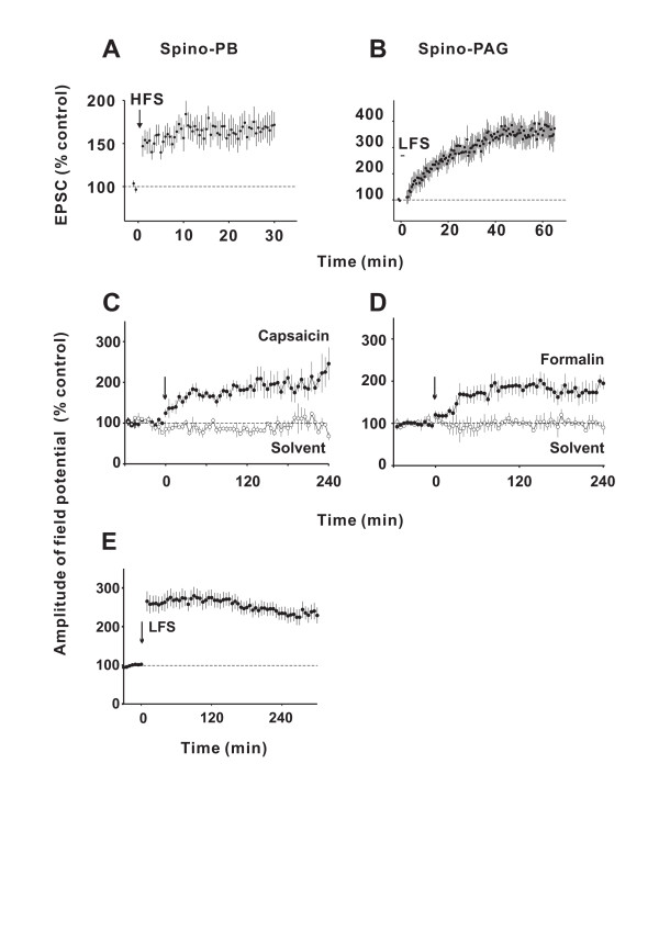
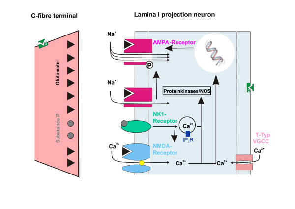
Long-term potentiation (LTP) at synapses of nociceptive nerve fibres is a proposed cellular mechanism underlying some forms of hyperalgesia. In this review fundamental properties of LTP in nociceptive pathways are described. The following topics are specifically addressed: A concise definition of LTP is given and a differentiation is made between LTP and "central sensitisation". How to (and how not to) measure and how to induce LTP in pain pathways is specified. The signal transduction pathways leading to LTP at C-fibre synapses are highlighted and means of how to preempt and how to reverse LTP are delineated. The potential functional roles of LTP are evaluated at the cellular level and at the behavioural level in experimental animals. Finally, the impact of LTP on the perception of pain in human subjects is discussed.


Figures

Figure 1
Figure 1
A, B show contrasting forms of LTP expressed in distinct groups of spinal lamina I projection neurons in vitro. Time courses of mean amplitudes (± SEM) of C-fibre-evoked EPSCs in lamina I neurons with a projection to the parabrachial area (PB, n = 8) or the periaqueductal grey (PAG, n = 7). Conditioning HFS induced LTP in all spino-PB neurons tested (A) but was ineffective in spino-PAG neurons (data not shown). Conditioning LFS induced LTP in all 18 spino-PAG neurons tested (B) but was never effective in seven spino-PB neurons (data not shown). C-E LTP can be induced by natural, low frequency afferent barrage evoked by inflammation of peripheral tissue in vivo. Mean time courses of C-fibre-evoked field potentials recorded extracellulary in superficial spinal dorsal horn in response to electrical stimulation of left sciatic nerve of deeply anaesthetized adult rats with spinal cords and afferent nerves intact. Subcutaneous injections of transient receptor potential vanilloid 1 channel agonist capsaicin (1%, 100 μl, n = 5, C) or formalin (5%, 100 μl, n = 6, D) into the glabrous skin at the ipsilateral hind paw, within the innervation territory of the sciatic nerve at time zero (arrows) induced LTP (closed circles), while injections of the respective solvents (open circles) had no effects (n = 3 in each group). Conditioning electrical LFS (2 Hz, 2 min at C-fibre intensity) of sciatic nerve at time zero (arrow) induced LTP (n = 28, E) which was prevented by NMDA receptor antagonist MK-801 (3 mg kg-1, i.v.-infusion over 30 min, data not shown). Modified from [12].
Figure 2
Figure 2
Potential mechanisms of potentiation, prevention and de-potentiation at synapses between C-fibres and spinal cord projection neurons. Conditioning electrical nerve stimulation or natural noxious stimulation triggers release of glutamate and substance P which causes opening of NMDA receptor channels and T-type voltage-gated Ca2+ channel and Ca2+ release from intracellular stores. This activates Ca2+-dependent signal transduction pathways including protein kinases and transcription factors. Synaptic strength is probably increased by phosphorylation of synaptic proteins including AMPA receptor channels [55], altered trafficking of synaptic proteins, e.g. increased insertion of AMPA receptors into the sub-synaptic membrane [56] and de-novo protein synthesis. According to this model, LTP can be prevented if release of glutamate and/or substance P is inhibited, for example by activation of pre-synaptic, G-protein-coupled μ-opioid receptors, or if opening of voltage sensitive and Ca2+ permeable ion channels is blocked, e.g. via postsynaptic inhibition by an opioid. Depotentiation could result from de-phosphorylation of synaptic proteins, changes in receptors trafficking and degradation of synaptic proteins.

References

    1. Bliss TVP, Collingridge GL. A synaptic model of memory: long-term potentiation in the hippocampus. Nature. 1993;361:31–39. doi: 10.1038/361031a0. - DOI - PubMed
    1. Malenka RC, Bear MF. LTP and LTD: an embarrassment of riches. Neuron. 2004;44:5–21. doi: 10.1016/j.neuron.2004.09.012. - DOI - PubMed
    1. Bailey CH, Kandel ER, Si K. The persistence of long-term memory: a molecular approach to self-sustaining changes in learning-induced synaptic growth. Neuron. 2004;44:49–57. doi: 10.1016/j.neuron.2004.09.017. - DOI - PubMed
    1. Lisman J, Raghavachari S. A unified model of the presynaptic and postsynaptic changes during LTP at CA1 synapses. Sci STKE. 2006;2006:1–15. - PubMed
    1. Mantyh PW, Rogers SD, Honoré P, Allen BJ, Ghilardi JR, Li J, et al. Inhibition of hyperalgesia by ablation of lamina I spinal neurons expressing the substance P receptor. Science. 1997;278:275–279. doi: 10.1126/science.278.5336.275. - DOI - PubMed

Publication types