. 2007 Apr 3:8:92.
doi: 10.1186/1471-2164-8-92.

Human sterile alpha motif domain 9, a novel gene identified as down-regulated in aggressive fibromatosis, is absent in the mouse

Affiliations

Human sterile alpha motif domain 9, a novel gene identified as down-regulated in aggressive fibromatosis, is absent in the mouse

Catherine F Li et al. BMC Genomics. .

Abstract

Background: Neoplasia can be driven by mutations resulting in dysregulation of transcription. In the mesenchymal neoplasm, aggressive fibromatosis, subtractive hybridization identified sterile alpha motif domain 9 (SAMD9) as a substantially down regulated gene in neoplasia. SAMD9 was recently found to be mutated in normophosphatemic familial tumoral calcinosis. In this study, we studied the gene structure and function of SAMD9, and its paralogous gene, SAMD9L, and examined these in a variety of species.

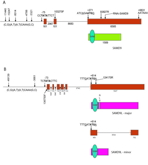
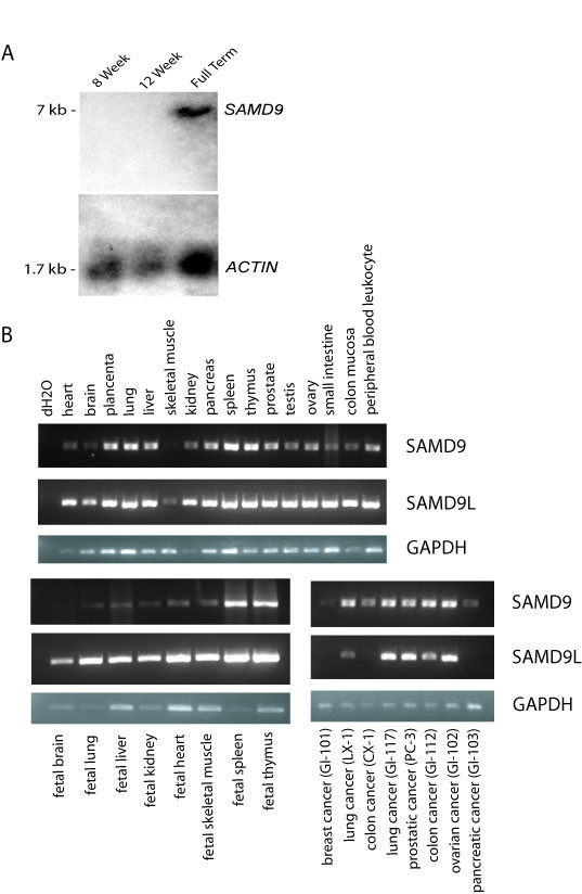
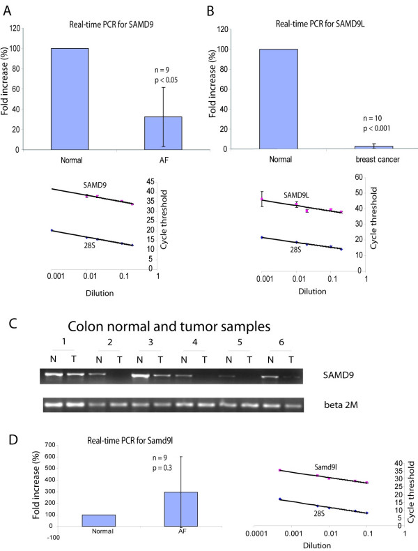
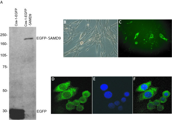
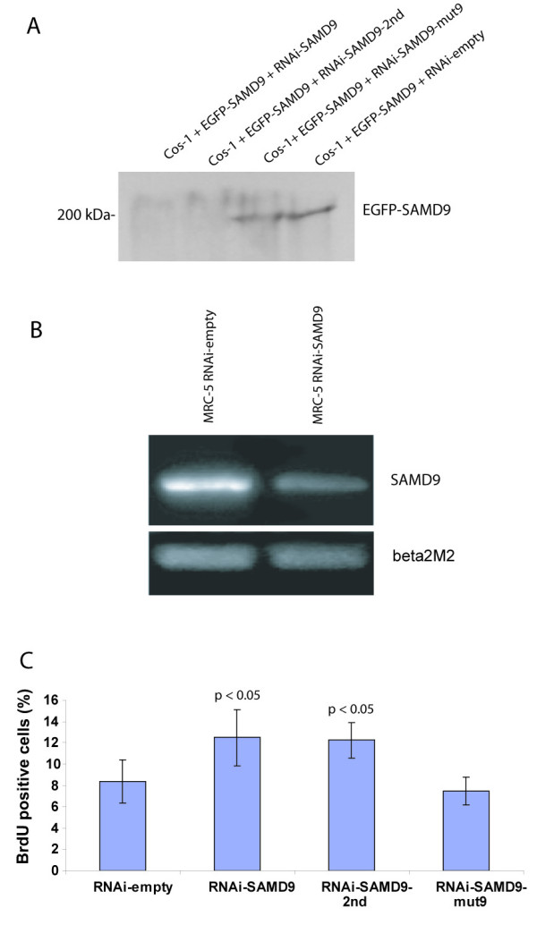
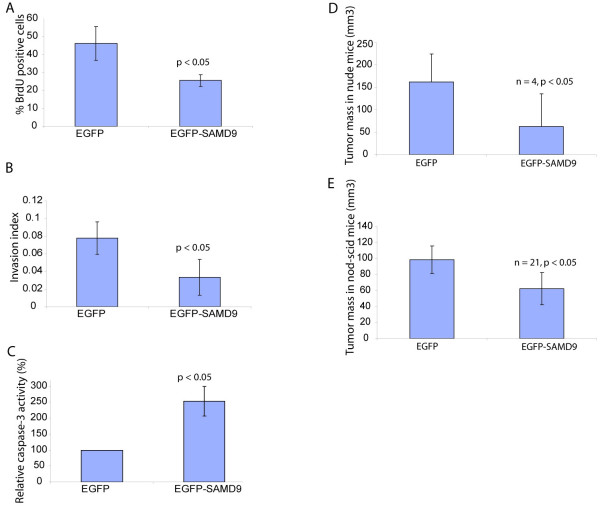
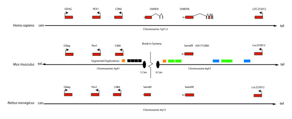
Results: SAMD9 is located on human chromosome 7q21.2 with a paralogous gene sterile alpha motif domain 9 like (SAMD9L) in the head-to-tail orientation. Although both genes are present in a variety of species, the orthologue for SAMD9 is lost in the mouse lineage due to a unique genomic rearrangement. Both SAMD9 and SAMD9L are ubiquitously expressed in human tissues. SAMD9 is expressed at a lower level in a variety of neoplasms associated with beta-catenin stabilization, such as aggressive fibromatosis, breast, and colon cancers. SAMD9 and SAMD9L contain an amino-terminal SAM domain, but the remainder of the predicted protein structure does not exhibit substantial homology to other known protein motifs. The putative protein product of SAMD9 localizes to the cytoplasm. In vitro data shows that SAMD9 negatively regulates cell proliferation. Over expression of SAMD9 in the colon cancer cell line, SW480, reduces the volume of tumors formed when transplanted into immune-deficient mice.

Conclusion: SAMD9 and SAMD9L are a novel family of genes, which play a role regulating cell proliferation and suppressing the neoplastic phenotype. This is the first report as far as we know about a human gene that exists in rat, but is lost in mouse, due to a mouse specific rearrangement, resulting in the loss of the SAMD9 gene.


Figures

Figure 1
Figure 1
Gene and protein structure of SAMD9 and SAMD9L. A. Diagram of SAMD9 gene structure and predicted protein structure. Exons are shown as red rectangles. The protein is shown in green, with the sterile alpha motif domain as blue oval. B. Diagram of SAMD9L gene structure and predicted protein structure. Exons are shown in red. The protein is shown as a pink box, with two predicted open reading frames of shown. The sterile alpha motif domain is shown as blue oval. A potential nuclear localization domain is shown as grey box.
Figure 2
Figure 2
Expression of SAMD9 and SAMD9L in multiple tissue types. A. Northern blot from total RNA of 8 weeks, 12 weeks and full term human placenta probed with 3'UTR of SAMD9. A 7 kb transcript was detected in full term human placenta. B. Human adult, fetal, and tumor multiple tissue cDNA panels were amplified with the sequences at 3'UTR of SAMD9, SAMD9L or GAPDH as primers. SAMD9 was expressed in all human adult, fetal and tumor tissues that were available from human multiple tissue cDNA panels except for fetal brain and breast cancer. It was expressed at a very low level in skeletal muscle and pancreatic cancer. SAMD9L is expressed in all human adult, fetal and tumor tissues that were available from human multiple tissue cDNA panels, except breast cancer, colon cancer, and pancreatic cancer.
Figure 3
Figure 3
Lower level of expression of SAMD9 and SAMD9L in neoplasia. A. Validation curves for the primer sets of SAMD9 and 28S rRNA, and real time quantitative PCR of SAMD9. SAMD9 and 28S rRNA have similar amplification efficiency since the slope was very similar for both curves. SAMD9 was expressed at a lower level in aggressive fibromatosis (AF) tumors compared to normal fibrous tissues from same patients. B. Validation curves for the primer sets of SAMD9L and 28S rRNA, and real time quantitative PCR of SAMD9L. SAMD9L and 28S rRNA have similar amplification efficiency since the slope was very similar for both curves. SAMD9L was expressed at a lower level in breast cancers compared to normal breast epithelial tissues from same patients.C. Semi-quantitative RT-PCR of SAMD9. SAMD9 was under-expressed in some cases of colon cancer compared to the normal colon mucosa from same patients. Real time PCR data showed no significant difference in expression level between breast and colon cancer and normal controls as a group. D. There was no significant difference in expression of SAMD9L between aggressive fibromatosis samples and normal controls tissues.
Figure 4
Figure 4
SAMD9 protein is expressed and localized to the cytoplasm. A. Western analysis for the SAMD9-EGFP fusion protein. The SAMD9 fusion protein was detected as a predicted 200 kDa band in Cos-1 cells, using an antibody to GFP. B, C. Expression of SAMD9 putative protein in MRC-5 cells. N-terminal EGFP tagged SAMD9 was detected in the cytoplasm of MRC-5 cells when transiently transfected. B. Light field view. C. Fluorescent microscopy view for FITC. D, E, F. Expression of SAMD9 putative protein in SW480 cells. N-terminal EGFP tagged SAMD9 localized to the cytoplasm of SW480 cells. D. Fusion protein detected under fluorescent wave length for FITC. E. Cell nucleus detected under fluorescent wave length for DAPI. F. Merged view of image D and E.
Figure 5
Figure 5
SAMD9 regulates cell proliferation. A. RNAi for SAMD9 is effective in reducing protein expression. A 200 kDa band for the SAMD9 fusion protein was detected in Cos-1 cells after co-transfection of pLP-EGFP-SAMD9 with either pSUPER-RNAi-SAMD9-mut9 or pSUPER-RNAi-EGFP, but it is not detected after co-transfection with either pSUPER-RNAi-SAMD9 or pSUPER-RNAi-SAMD9-2nd. The expression of the EGFP-SAMD9 fusion protein was reduced in Cos-1 cells with RNA interference of SAMD9. B. Semi-quantitative RT-PCR for SAMD9 expression in MRC-5 cells. SAMD9 expression was reduced after RNA interference. C. Proliferation rate (Brdu incorporation percent) increased after RNA interference of SAMD9 in MRC-5 cells.
Figure 6
Figure 6
SAMD9 reduces tumor size. A. Increased expression of SAMD9 reduced SW480 cell proliferation rate as detected by Brdu incorporation percent. B. Increased expression of SAMD9 reduced the invasion index of SW480 cells. C. Increased expression of SAMD9 increased caspase activity in SW480 cells. D and E. The tumor volume formed from SW480 xenografts was reduced with increased expression of SAMD9 when transplanted into nude mice (D) or Nod-scid mice (E).
Figure 7
Figure 7
Diagram for the genomic region encompassing the loci of SAMD9 and SAMD9L in human, mouse and rat along with the segmental duplications. Exons are shown as a red box. Segmental duplications are shown as orange, green, blue and grey boxes. Two orange segmental duplications may mediate chromosomal breakage with subsequent deletion of SAMD9, and the centromeres of chromosome 6 and 5 are formed at the sites of breakage. The grey segmental duplications, sharing the sequence among numerous centromeric loci, are accumulated at the centromere of chromosome 5. The green and blue segmental duplications flanking the SAMD9L are mouse specific based on sequence similarity, while the orange and grey segments have occurred at about the same time as the divergence of mouse and rat.

Similar articles

Cited by

References

    1. Martin B, Schneider R, Janetzky S, Waibler Z, Pandur P, Kuhl M, Behrens J, von der Mark K, Starzinski-Powitz A, Wixler V. The LIM-only protein FHL2 interacts with beta-catenin and promotes differentiation of mouse myoblasts. J Cell Biol. 2002;159:113–122. doi: 10.1083/jcb.200202075. - DOI - PMC - PubMed
    1. Goruppi S, Chiaruttini C, Ruaro ME, Varnum B, Schneider C. Gas6 induces growth, beta-catenin stabilization, and T-cell factor transcriptional activation in contact-inhibited C57 mammary cells. Mol Cell Biol. 2001;21:902–915. doi: 10.1128/MCB.21.3.902-915.2001. - DOI - PMC - PubMed
    1. Alman BA, Pajerski ME, Diaz-Cano S, Corboy K, Wolfe HJ. Aggressive fibromatosis (desmoid tumor) is a monoclonal disorder. Diagn Mol Pathol. 1997;6:98–101. doi: 10.1097/00019606-199704000-00005. - DOI - PubMed
    1. Li C, Bapat B, Alman BA. Adenomatous polyposis coli gene mutation alters proliferation through its beta-catenin-regulatory function in aggressive fibromatosis (desmoid tumor) Am J Pathol. 1998;153:709–714. - PMC - PubMed
    1. Tejpar S, Li C, Yu C, Poon R, Denys H, Sciot R, Van Cutsem E, Cassiman JJ, Alman BA. Tcf-3 expression and beta-catenin mediated transcriptional activation in aggressive fibromatosis (desmoid tumour) Br J Cancer. 2001;85:98–101. doi: 10.1054/bjoc.2001.1857. - DOI - PMC - PubMed

Publication types

MeSH terms

LinkOut - more resources