. 2007 Mar;25(6):1705-10.
doi: 10.1111/j.1460-9568.2007.05454.x. Epub 2007 Apr 4.

Proteolytic cleavage of the voltage-gated Ca2+ channel alpha2delta subunit: structural and functional features

Affiliations

Proteolytic cleavage of the voltage-gated Ca2+ channel alpha2delta subunit: structural and functional features

Arturo Andrade et al. Eur J Neurosci. 2007 Mar.

Abstract

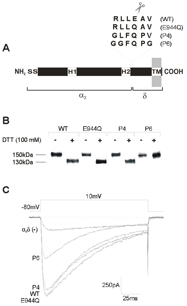
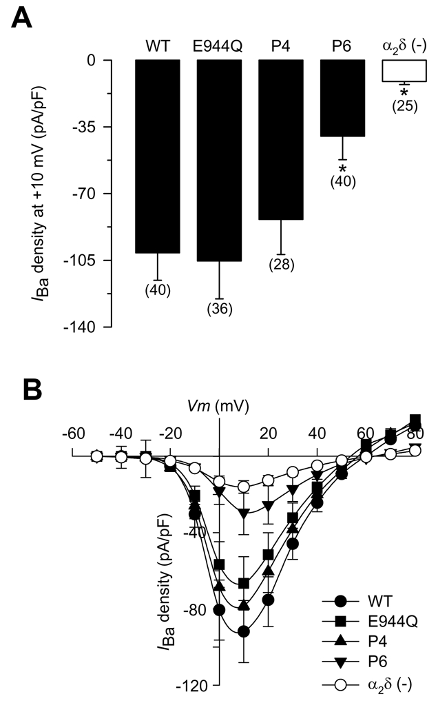
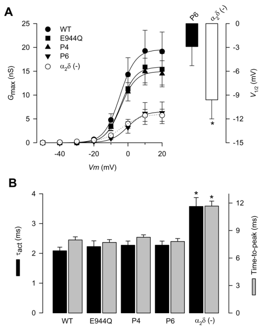
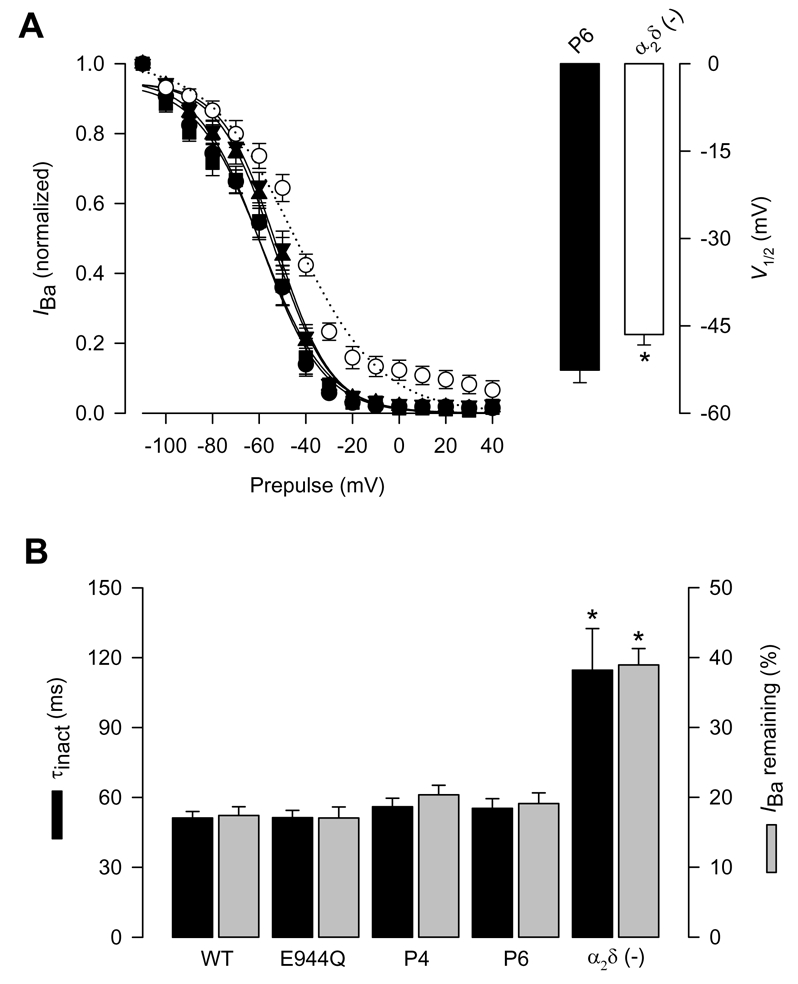
By mediating depolarization-induced Ca(2+) influx, high-voltage-activated Ca(2+) channels control a variety of cellular events. These heteromultimeric proteins are composed of an ion-conducting (alpha(1)) and three auxiliary (alpha(2)delta, beta and gamma) subunits. The alpha(2)delta subunit enhances the trafficking of the channel complex to the cell surface and increases channel open probability. To exert these effects, alpha(2)delta must undergo important post-translational modifications, including a proteolytic cleavage that separates the extracellular alpha(2) from its transmembrane delta domain. After this proteolysis both domains remain linked by disulfide bonds. In spite of its central role in determining the final conformation of the fully mature alpha(2)delta, almost nothing is known about the physiological implications of this structural modification. In the current report, by using site-directed mutagenesis, the proteolytic site of alpha(2)delta was mapped to amino acid residues Arg-941 and Val-946. Substitution of these residues renders the protein insensitive to proteolytic cleavage as evidenced by the lack of molecular weight shift upon treatment with a disulfide-reducing agent. Interestingly, these mutations significantly decreased whole-cell patch-clamp currents without affecting the voltage dependence or kinetics of the channels, suggesting a reduction in the number of channels targeted to the plasma membrane.


Figures

Figure 1
Figure 1. Expression of the α2δ subunit proteolytic site mutants in HEK-293 cells
A) Schematic representation of the CaVα2δ auxiliary subunit. The location of the two hydrophobic domains (H1 and H2), and the signal sequence (S) are given. The amino acid residues in the putative proteolytic site of the wild-type α2δ-1 and its mutant versions are aligned and shown above the diagram. B) Western blot analysis of membranes from HEK-293 cells expressing the wild-type, and the mutant versions of the α2δ subunit (E944Q, P4 and P6). Membranes were prepared as described under Material and Methods. Only the sextuple mutation (P6) is insensitive to proteolysis (do not change its electrophoretic pattern after dithiothreitol-DDT-treatment). C) Representative Ba2+ currents through recombinant CaV2.2/β3 channels obtained in HEK-293 cells co-expressing the wild-type α2δ-1 and its proteolysis mutants. Currents were elicited in response to 140 ms test pulses to +10 mV delivered from a holding potential of −80 mV.
Figure 2
Figure 2. Functional effects of heterologous expression of the wild-type α2δ subunit and its proteolytic site mutants
A) Average ± SEM IBa density in HEK-293 cells expressing the wild-type α2δ-1 and its mutant variants (filled bars). IBa density was calculated from IBa amplitude at a test pulse of +10 mV normalized to cell membrane capacitance. The open bar shows the average current amplitude in the absence of the α2δ subunit. The number of recorded cells is indicated in parentheses, and the asterisks denote significant differences (P<0.05). B) Average ± SEM I–V relationships for IBa recorded from HEK-293 cells expressing the wild-type α2δ-1 and its mutant variants (E944Q, P4 and P6). IBa density was calculated at a series of test pulses applied from a holding potential of −80 mV in 10 mV steps between −50 and +80 mV (n = 6–8). Open circles represent the average current density in the absence of the α2δ subunit.
Figure 3
Figure 3. Activation properties of CaV2.2/β3 channels co-expressed with the wild-type α2δ-1or its proteolysis mutations
A) Left, voltage dependence of activation of CaV2.2/β3 channels co-expressed with wild-type α2δ-1 (filled circles), its proteolysis mutants (E944Q, P4 and P6) or without any α2δ subunit (open circles). Maximum conductance (Gmax) plotted data were derived from Fig. 2B. The mean data were fitted with Boltzmann functions, the V½ values of which are given in Table 1. Right, comparison of the mean half-activation voltage (V1/2) for CaV2.2/β3 channels co-expressed with the P6 proteolysis mutation or without the α2δ subunit (n = 6–8). B) Mean time to peak and time constants of activation (τact) obtained by fitting the rising phase of IBa at +10 mV with a single exponential, for CaV2.2/β3 channels co-expressed with wild-type α2δ-1, its proteolysis mutants, or without any α2δ subunit (n = 11–25).
Figure 4
Figure 4. Inactivation properties of CaV2.2/β3 channels co-expressed with the wild-type α2δ-1 or its proteolysis mutations
A) Left, voltage dependence of steady-state inactivation of CaV2.2/β3 channels co-expressed with wild-type CaVα2δ-1 (filled circles), its proteolysis mutants E944Q, P4 and P6 or without any α2δ subunit (open circles). Currents were recorded after conditioning pulses of 1 s duration, applied from a holding potential of −80 mV in 10 mV steps between −110 and +40 mV, followed by a 140 ms test pulse to +10 mV. The normalized data are plotted against the conditioning potentials (n = 5–11). The mean data were fitted with a Boltzmann function, the V½ values of which are given in Table 1. Right, comparison of the mean half-inactivation voltage (V1/2) for CaV2.2/β3 channels co-expressed with the P6 mutation or without the α2δ subunit. B) Average time constants of inactivation (τinact) and percentage of current remaining 140 ms into the depolarizing pulse for CaV2.2/β3 channels co-expressed with wild-type α2δ-1, its proteolysis mutants, or without any α2δ subunit (n = 11–25).

References

    1. Arikkath J, Campbell KP. Auxiliary subunits: essential components of the voltage-gated calcium channel complex. Curr Opin Neurobiol. 2003;13:298–307. - PubMed
    1. Avila G, Sandoval A, Felix R. Intramembrane charge movement associated with endogenous K+ channel activity in HEK-293 cells. Cell Mol Neurobiol. 2004;24:317–330. - PMC - PubMed
    1. Bangalore R, Mehrke G, Gingrich K, Hofmann F, Kass RS. Influence of L-type Ca channel α2δ subunit on ionic and gating current in transiently transfected HEK 293 cells. Am J Physiol. 1996;270:H1521–H528. - PubMed
    1. Barclay J, Balaguero N, Mione M, Ackerman SL, Letts VA, Brodbeck J, Canti C, Meir A, Page KM, Kusumi K, PerezReyes E, Lander ES, Frankel WN, Gardiner RM, Dolphin AC, Rees M. Ducky mouse phenotype of epilepsy and ataxia is associated with mutations in the Cacna2d2 gene and decreased calcium channel current in cerebellar Purkinje cells. J Neurosci. 2001;21:6095–6104. - PMC - PubMed
    1. Bernstein GM, Jones OT. Kinetics of internalization and degradation of N-type voltage-gated calcium channels: Role of the α2/δ subunit. Cell Calcium. 2006 (in press) - PubMed

Publication types

Substances