. 2007 Apr 4:7:26.
doi: 10.1186/1471-2180-7-26.

Genetic microheterogeneity and phenotypic variation of Helicobacter pylori arginase in clinical isolates

Affiliations

Genetic microheterogeneity and phenotypic variation of Helicobacter pylori arginase in clinical isolates

Justin G Hovey et al. BMC Microbiol. .

Abstract

Background: Clinical isolates of the gastric pathogen Helicobacter pylori display a high level of genetic macro- and microheterogeneity, featuring a panmictic, rather than clonal structure. The ability of H. pylori to survive the stomach acid is due, in part, to the arginase-urease enzyme system. Arginase (RocF) hydrolyzes L-arginine to L-ornithine and urea, and urease hydrolyzes urea to carbon dioxide and ammonium, which can neutralize acid.

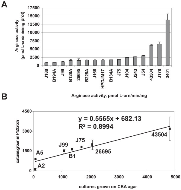
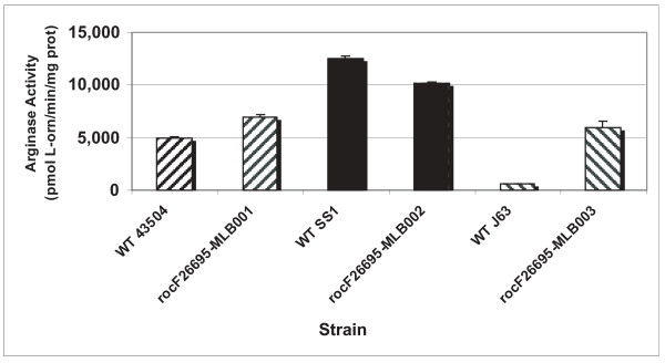
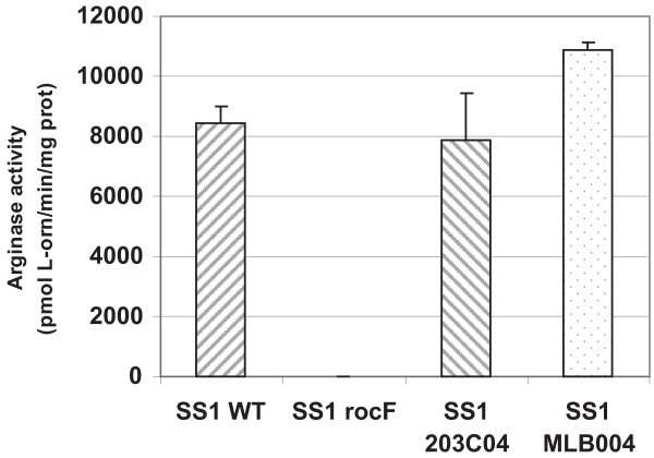
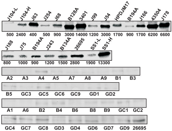
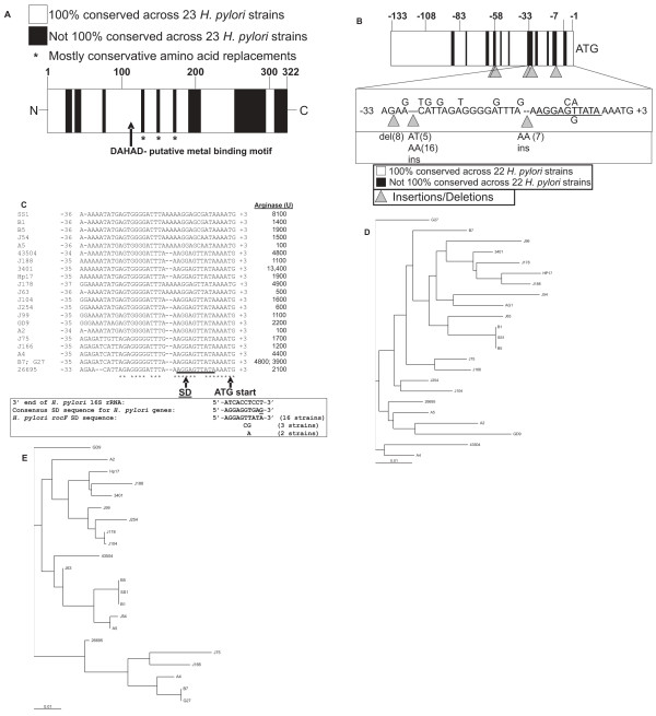
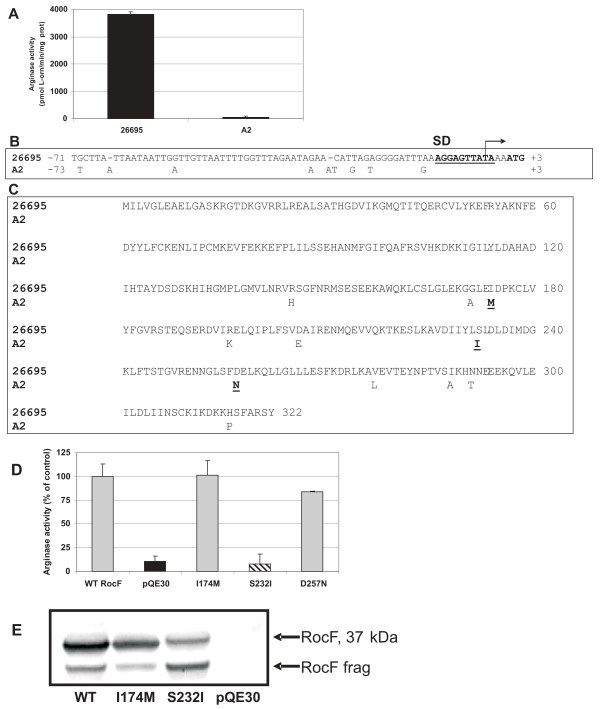
Results: The degree of variation in arginase was explored at the DNA sequence, enzyme activity and protein expression levels. To this end, arginase activity was measured from 73 minimally-passaged clinical isolates and six laboratory-adapted strains of H. pylori. The rocF gene from 21 of the strains was cloned into genetically stable E. coli and the enzyme activities measured. Arginase activity was found to substantially vary (>100-fold) in both different H. pylori strains and in the E. coli model. Western blot analysis revealed a positive correlation between activity and amount of protein expressed in most H. pylori strains. Several H. pylori strains featured altered arginase activity upon in vitro passage. Pairwise alignments of the 21 rocF genes plus strain J99 revealed extensive microheterogeneity in the promoter region and 3' end of the rocF coding region. Amino acid S232, which was I232 in the arginase-negative clinical strain A2, was critical for arginase activity.

Conclusion: These studies demonstrated that H. pylori arginase exhibits extensive genotypic and phenotypic variation which may be used to understand mechanisms of microheterogeneity in H. pylori.


Figures

Figure 1
Figure 1
Arginase activity variation in H. pylori clinical isolates. Ornithine concentration was measured at 515 nm spectrophotometerically by the appearance of an orange color originating from the reaction of ornithine with acidified ninhydrin. A. Arginase activity of extracts from 16 US H. pylori strains, 11 of which are minimally-passaged clinical H. pylori isolates. Strains were grown on Campylobacter blood agar plates for 48 h. The graphs show the average arginase activity (pmol L-Orn/min/mg protein) ± standard deviation of one experiment representative of at least three. A complete list of strains and average arginase activities is given in Table S1 (see Additional file 1). The variation from experiment to experiment is about 10–15%. B. Comparison of arginase activity of H. pylori grown in broth versus grown on agar. H. pylori strains were grown in Ham's F-12 broth for 16–18 h or on Campylobacter blood agar plates for 48 h as described in Materials and Methods. The data suggest a strong correlation between the arginase activity from H. pylori grown in broth versus that on agar.
Figure 2
Figure 2
Strain-specific regulation of H. pylori arginase activity. Comparison of arginase activities of a native H. pylori strain and a rocF mutant strain of 26695 complemented with the corresponding rocF gene (coding region plus native rocF promoter). Strain rocF-26695-MLB001 is a rocF mutant of strain 26695 in which the wild type rocF gene from strain 43504 has been complemented. The MLB002 strain carried wild type rocF from strain SS1 and the MLB003 strain carried wild type rocF from strain J63.
Figure 3
Figure 3
H. pylori prevents arginase activity from becoming too elevated. Wild type H. pylori strain SS1 was transformed with the rocF mutation (rocF), a vector control inserted into the intergenic hp0203-0204 site (203C04), or with the intergenic plasmid carrying the wild type rocF coding region and native promoter (MLB004).
Figure 4
Figure 4
Anti-RocF Western on minimally-passaged wild type H. pylori. Proteins were separated by SDS-PAGE using a 12% resolving gel and 10 μg of protein loaded per lane and analyzed by anti-RocF antibodies. Only a subset of strains is shown. J104-L, low arginase activity strain minimally passaged; J104-H, high arginase activity strain passaged for seven to nine days; SS1-L, low arginase activity strain passaged for seven to nine days; SS1-H, high arginase activity strain minimally-passaged. Numbers below the top two blots represent the arginase specific activity (rounded to nearest 100; pmol L-ornithine/min/mg protein ± standard deviation) of the extract used for the blot.
Figure 5
Figure 5
Microheterogeneity in arginase sequences from clinical isolates of H. pylori. The arginase coding region (~1.0 kb) plus ~130 bp of the upstream region encompassing the promoter and 18 bp downstream were amplified by PCR using Pfx and cloned into pBluescript. The constructs were sequenced using T3 and T7-1 primers (see Materials and Methods). A. Summary of microheterogeneity in the arginase coding region in H. pylori. Multi-sequence pairwise alignments were conducted with the rocF coding region using ClustalW. The alignment was converted to a graphical representation to show regions that are 100% conserved (white) and regions that vary (black). B. Hypervariablity in the ~133 bp rocF upstream region including the arginase promoter sequence preceding the ATG start codon. Multi-sequence pairwise alignments of the ~133 bp rocF upstream region from H. pylori strains were conducted using ClustalW. The alignments were converted to a graphical representation to show regions that are 100% conserved (white) and regions that vary (black). The alignments revealed a hypervariable region proximal to the ATG start codon. Numbering based on that of H. pylori strain 26695. Underlined is the Shine-Dalgarno (SD) sequence. Numbers in parentheses correspond to the number of strains featuring the insertion (ins) or deletion (del). Strain AG1 was omitted from this analysis because its arginase upstream region was completely different from the other 22 strains. C. Hypervariability in the ~35 bp upstream of the arginase ATG translation start codon in 21 H. pylori strains. Some of the sequence hypervariability occurred in the predicted SD region (underlined). The consensus sequence for the 3' end of the 16S rRNA for H. pylori is shown, as well as the consensus SD sequence for other H. pylori genes. Arginase activity is shown on the right hand side in units rounded to the nearest 100. Asterisks indicate nucleotides conserved in all strains. D. Phylogram of RocF protein sequence from 23 H. pylori strains. ClustalX and Treeview were used to construct the tree. E. Phylogram of the rocF upstream region from 22 H. pylori strains. ClustalX and Treeview were used to construct the tree. Strain AG1 was omitted from this analysis because its arginase upstream region was completely different from the other 22 strains.
Figure 6
Figure 6
Molecular basis of the arginase null phenotype of H. pylori clinical isolate A2. A. Arginase activity of H. pylori strain 26695 and clinical isolate A2. B. Variation in the arginase upstream regions of H. pylori strains 26695 and A2. Nine nucleotide differences were noted between these two sequences. C. Variations in the arginase amino acid sequences between H. pylori strains 26695 and A2. Eleven amino acid differences are highlighted (311/322 identity [96% identity]; 316/322 similar [97% similar]. Three amino acid differences (underlined) were found only in strain A2: I174M, S232I, and D257N. D. Arginase activity of E. coli transformed with pQE30-rocF (rocF from strain 26695) and of strains transformed with site-directed mutants of I174M, S232I, and D257N. E. Anti-RocF Western blot analysis of extracts of E. coli transformed with different plasmids including site-directed mutants of RocF. RocF frag indicates degradation product from RocF.

Similar articles

Cited by

References

    1. Blaser MJ. Gastric Campylobacter-like organisms, gastritis, and peptic ulcer disease. Gastroenterology. 1987;93:371–383. - PubMed
    1. Blaser MJ. Helicobacter pylori and the pathogenesis of gastroduodenal inflammation. Journal of Infectious Diseases. 1990;161:626–633. - PubMed
    1. Marshall BJ, McGechie DB, Rogers PA, Glancy RJ. Pyloric Campylobacter infection and gastroduodenal disease. Medical Journal of Australia. 1985;142:439–444. - PubMed
    1. Nomura A, Stemmermann GN, Chyou PH, Kato I, Perez-Perez GI, Blaser MJ. Helicobacter pylori infection and gastric carcinoma among Japanese Americans in Hawaii.[see comment] New England Journal of Medicine. 1991;325:1132–1136. - PubMed
    1. Parsonnet J, Friedman GD, Vandersteen DP, Chang Y, Vogelman JH, Orentreich N, Sibley RK. Helicobacter pylori infection and the risk of gastric carcinoma. N Engl J Med. 1991;325:1127–1131. - PubMed

Publication types

MeSH terms

LinkOut - more resources