. 2007 May 25;146(3):1117-27.
doi: 10.1016/j.neuroscience.2007.02.006. Epub 2007 Apr 6.

Exploration of prostanoid receptor subtype regulating estradiol and prostaglandin E2 induction of spinophilin in developing preoptic area neurons

Affiliations

Exploration of prostanoid receptor subtype regulating estradiol and prostaglandin E2 induction of spinophilin in developing preoptic area neurons

S R Burks et al. Neuroscience. .

Abstract

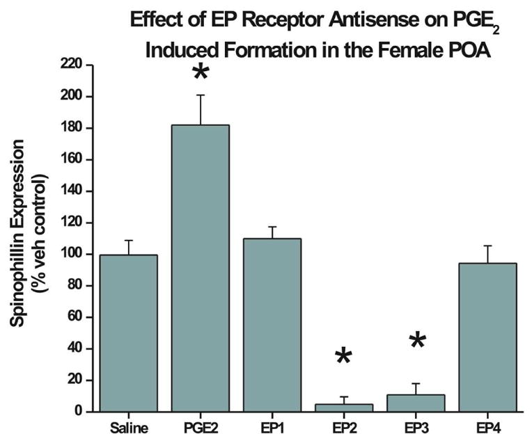
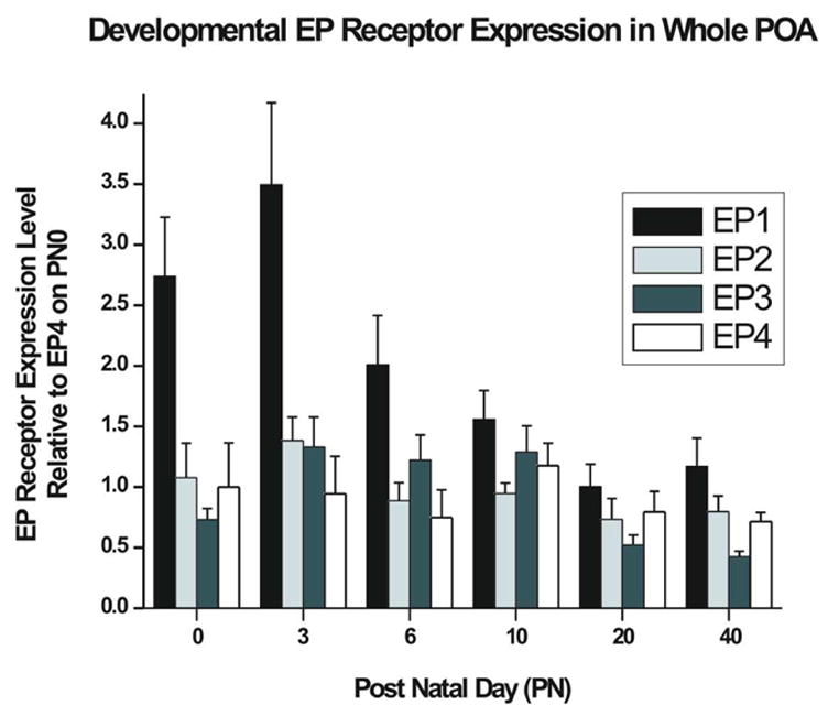
The prostaglandin E2 (PGE2) mediates estradiol-induced masculinization of sexual behavior in the rat during a perinatal sensitive period. PGE2 induces formation of dendritic spines on preoptic area (POA) neurons and this synaptic pattern change is associated with the ability to express male sexual behavior as an adult. Whether PGE2 is released from astrocytes or neurons in the developing POA is unknown. To further understanding of how PGE2 induces dendritic spine formation at the cellular level, we have explored the PGE2 receptor subtype mediating this response. There are four receptors for PGE2, EP1, EP2, EP3 and EP4, each having unique but interacting signal transduction profiles. Treatment of newborn female rats with the EP receptor agonists iloprost, butaprost and sulprostone indicated that stimulation of both the EP2 and EP3 receptors significantly increased spinophilin, a protein whose levels positively correlate to the presence of dendritic spines and masculinization of the POA. Use of antisense oligonucleotides against the mRNA for each receptor reveals that either EP2 or EP3 receptor knockdown reduces spinophilin in PGE2- or estradiol-treated females, whereas reducing EP1 or EP4 receptor levels by the same means has a smaller but also significant effect. A developmental profile of EP receptor expression indicates EP1 in particular is elevated for the first few days of life, corresponding to the critical period for masculinization, whereas mRNA levels for the other three receptors remain relatively constant.


Figures

FIG 1
FIG 1. Treatment of animals with selective prostanoid receptor agonists induces spinophilin in the POA
A) PGE2 treatment of female pups on the day of birth and PN1 induces a significant 88.4% increase in spinophilin in the POA by PN2 compared to females treated with saline (ANOVA; p < 0.5). There was no significant effect on spinophillin levels of treatment with the EP1 selective agonist Iloprost, the EP2 selective agonist, Butaprost, or the non-selective EP1/EP3 agonist, Sulprostone. However, co-application of Butaprost and Sulprostone induced a significant 222.2% increase in spinophilin over those treated with saline (n=5–6/group; p < 0.05). B) Representative Western blot for spinophilin protein. Each lane represents POA tissue from one animal. Each blot included saline control animals for standardization.
FIG 2
FIG 2. EP receptor protein levels are reduced after treatment with antisense oligonucleotide sequences specific for each prostanoid receptor
Newborn pups were treated with 1μg of specific antisense (AS) oligonucleotide sequences or scrambled (SCRAM) oligonucleotide for two days and levels of receptor protein were measured on the third day. Measurements were normalized to control samples then compared against each other for significant differences A) Western Blot of EP receptors. Rows are different proteins being blotted for, columns represent specific sequence of antisense treatment. B) Treatment with the EP1 antisense oligonucleotide sequence reduced EP1 receptor protein by 21.9% 1 (n=4; ANOVA, p<0.01). C) Treatment with EP2 antisense oligo reduced protein levels for that receptor by 30.0% (n=5; ANOVA, p<0.04). D) Treatment with the EP3 sequence resulted in a 24.8% decrease in the EP3 receptor protein (n=5; ANOVA, p<0.01). E) Treatment with the EP4 sequence resulted in a 19.6% decrease in the EP4 receptor expression (n=4; ANOVA, p<0.01).
FIG 3
FIG 3. Antisense oligonucleotides against EP receptors block PGE2 induction of spinophilin
Treatment of newborn female pups with PGE2 + scrambled oligo (SCRAM) on PN0 and PN1 induces a significant increase in spinophilin by PN2 (n=4; ANOVA; p<0.05). Pretreatment with antisense oligo against either EP2 or EP3 receptor for 4 hrs prior to PGE2, reduced spinophilin levels to almost undetectable and significantly below SCRAM + saline treated controls (n=6 for both groups; ANOVA; p<0.01). Pretreatment with antisense oligo against the EP1 (n=6) or EP4 (n=5) receptors before PGE2 reduced spinophilin levels comparable to those seen in SCRAM + saline treated females.
FIG 4
FIG 4. Antisense oligonucleotides against EP receptors block the effects of estradiol on spinophilin
Treatment with estradiol + SCRAM induced a significant increase in spinophilin (n=4) compared to animals treated with vehicle plus scrambled oligo. Pretreatment with antisense oligos against either EP2 or EP3 receptor decreased spinophilin levels to almost undetectable (n=5 for both groups), while treatment with sequences against EP1 (n=4) or EP4 (n=6) produced levels of spinophilin comparable to treated with oil vehicle (control for estradiol) and SCRAM oligo (ANOVA; p < 0.01)
FIG 5
FIG 5. Antisense oligonucleotides against EP receptors do not inhibit estradiol induction of spinophilin in the hippocampus
Animals were treated with estradiol with and without specific antisense oligo sequences. Hippocampal tissue from the same animals as in Figure 2 were assayed for spinophilin expression. While estradiol treatment significantly increases spinophilin protein compared to the vehicle treatment group, the increase is not attenuated by the prior application of antisense oligonucleotides (all groups n=4, ANOVA, p<0.01).
FIG 6
FIG 6. Relative Expression of Prostanoid Receptors throughout development
Relative quantification of EP Receptor gene expression by RT-PCR show that EP1 mRNA is at significantly higher levels than the other EP receptors from PN0 through PN6 (For PN0, 3, 20, and 40, n=8 [4 male and 4 female] PN6, n=6 [2 male and 4 female] and PN10 n=6 [4 male and 2 female], ANOVA; p<0.05). This corresponds to the perinatal time period where the POA is sensitive to estradiol induced masculinization (data from males and females are combined for presentation).

References

    1. Adam M, Boie Y, Rushmore T, Muller G, Bastien L, Mckee K, Metters K, Abramovitz M. Cloning and expression of three isoforms of the human EP3 prostanoid receptor. FEBS Lett. 1994;338:170–174. - PubMed
    1. Amateau SK, Alt JJ, Stamps CL, McCarthy MM. Brain estradiol content in newborn rats: Sex differences, regional heterogeneity and possible de novo synthesis by the female telencephalon. Endocrinology. 2004;145:2906–2917. - PubMed
    1. Amateau SK, McCarthy MM. Sexual differentiation of astrocyte morphology in the developing rat preoptic area. J Neuroendocrinol. 2002a;14:904–910. - PubMed
    1. Amateau SK, McCarthy MM. A novel mechanism of dendritic spine plasticity involving estradiol induction of prostaglandin-E2. J Neurosci. 2002b;22:8586–8596. - PMC - PubMed
    1. Amateau S, McCarthy M. Induction of PGE2 by estradiol mediates developmental masculinization of sex behavior. Nat Neuroscience. 2004;7:643–650. - PubMed

MeSH terms

LinkOut - more resources