. 2007 May 25;146(3):1193-211.
doi: 10.1016/j.neuroscience.2007.01.072. Epub 2007 Apr 3.

Expression and immunolocalization of the plasma membrane monoamine transporter in the brain

Affiliations

Expression and immunolocalization of the plasma membrane monoamine transporter in the brain

A Dahlin et al. Neuroscience. .

Abstract

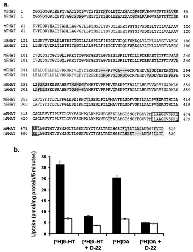
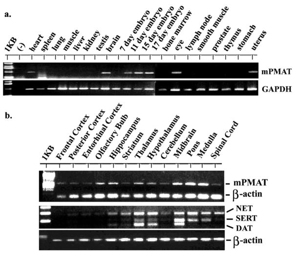
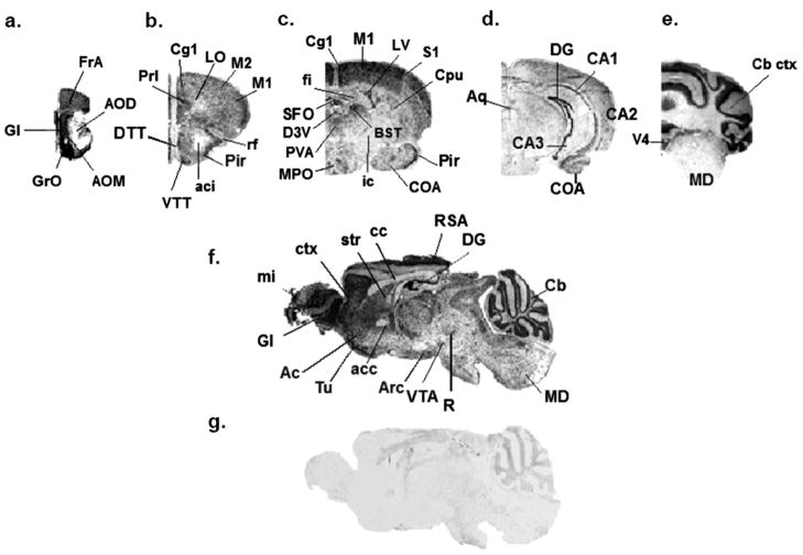
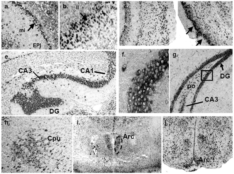
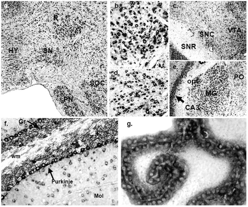
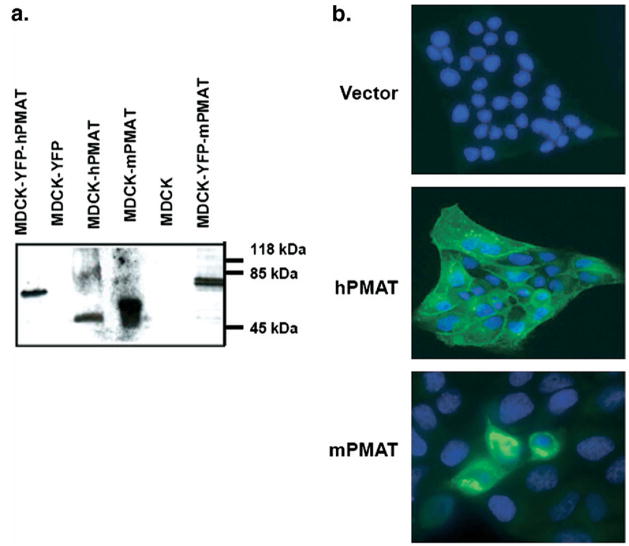
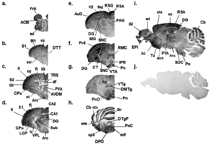
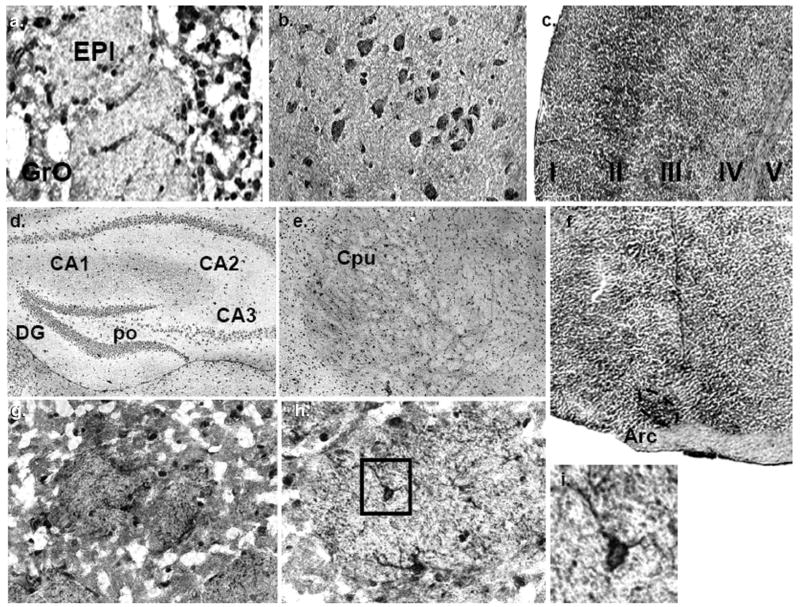
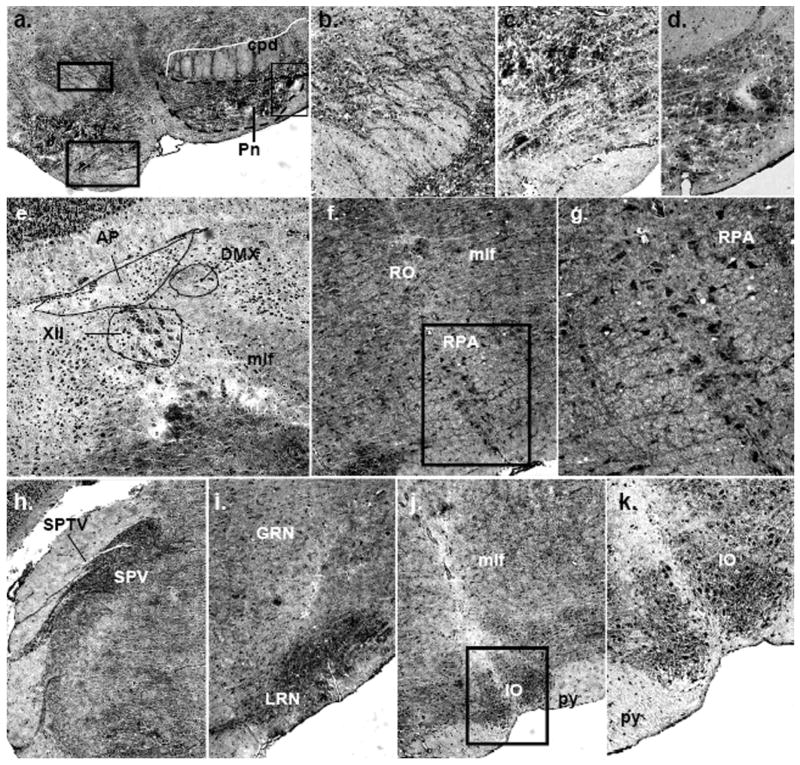
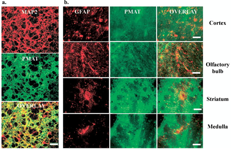
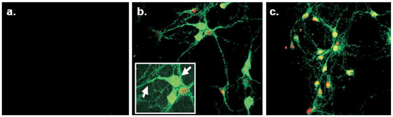
High affinity monoamine transporters efficiently terminate neurotransmission through synaptic reuptake of released neurotransmitter. We recently cloned and characterized a novel low-affinity, high capacity plasma membrane monoamine transporter (PMAT) that is strongly expressed in the human brain and efficiently transports 5-HT and dopamine (DA). In efforts to understand the physiological function of PMAT and its relevance in monoaminergic pathways, we cloned the PMAT homolog from the mouse brain, demonstrated its capability for transporting 5-HT and DA, and determined the regional and cellular localization of mouse plasma membrane monoamine transporter (mPMAT) in adult mouse brain by reverse-transcription polymerase chain reaction, non-radioactive in situ hybridization, and immunohistochemical methods. Our results showed that mPMAT mRNA and protein are broadly expressed in the mouse brain and are particularly abundant in forebrain cortex, olfactory tubercle, hippocampus, cerebellum and epithelial cells of the choroid plexus. Dual-immunofluorescence histochemistry with established phenotypic markers microtubule-associated protein (MAP2) and glial fibrillary acidic protein (GFAP) revealed that mPMAT is expressed in neuronal cells but not in astrocytes. mPMAT is co-expressed in many brain regions with the high affinity 5-HT transporter (SERT) and the dopamine transporter (DAT), but is also found in certain sites that receive monoamine innervation but lack significant expression of SERT or DAT. These findings suggest that mPMAT is a widely distributed, neuronally-expressed transporter, which may support the role of 5-HT and DA uptake under certain conditions.


Figures

Fig. 1
Fig. 1
Cloning and functional characterization of mPMAT. (a) Amino acid sequence alignment of mPMAT and hPMAT. Shaded areas correspond to non-conserved regions in the mPMAT and hPMAT amino acid sequences. The boxed region indicates the peptide sequence used to generate the p469 antibody. (b) Transport of DA and 5-HT (1 μM each) by MDCK cells stably transfected with either pEYFP-C1 vector (open bars) or mPMAT-pEYFP (filled bars), in the presence and absence of decynium-22 (D-22) (1 μM), a specific inhibitor of hPMAT.
Fig. 2
Fig. 2
Tissue and brain regional distribution of mPMAT mRNA analyzed by RT-PCR. (a) Tissue distribution of mPMAT analyzed by PCR using a mouse multi-tissue cDNA panel. (b) Top panel shows RT-PCR amplification of mPMAT transcripts from different brain regions. The bottom panel shows multiplex PCR analysis of mouse NET, SERT and DAT in various regions of the mouse brain. In all samples, amplification of GAPDH or β-actin was performed as an internal control.
Fig. 3
Fig. 3
Regional distribution of mPMAT mRNA in the mouse brain analyzed by non-radioactive ISH. Coronal (a–e) or sagittal (f–g) cryosections of mouse brain were subjected to non-radioactive DIG ISH using cRNA probes specific for mPMAT. Panels a–f show ISH labeling with the antisense probe whereas panel g represents the sense probe control. Images were obtained at 2× magnification.
Fig. 4
Fig. 4
Localization of mPMAT mRNA in forebrain regions as revealed by non-radioactive ISH. (a) mPMAT antisense cRNA probes densely label the mitral (mi) and glomerular layers (Gl) of the olfactory bulb. (b–d) Layers of the frontal cortex (b and c) and piriform cortex (d) are strongly labeled. (e–g) Intense labeling of mPMAT mRNA in the granular layers of the dentate gyrus (DG), CA1 and CA3 regions of the hippocampus. (f) High magnification (60×) of boxed area in panel g showing cytoplasmic staining of hippocampus granule cells. (h) Expression of mPMAT in the caudate putamen (striatum). (i, j) Expression of mPMAT in the arcuate nucleus (area surrounded by dashed line) of the hypothalamus.
Fig. 5
Fig. 5
Localization of mPMAT mRNA in midbrain and hindbrain as revealed by non-radioactive ISH. (a) mPMAT mRNA is observed in the midbrain substantia nigra (SN) and red nucleus (R), pons (PN and SOC) and hypothalamus (HY). The regions in panels b and d correspond to high-magnification (40×) images of the R and SN in the section from panel a. (c) mPMAT mRNA expression in the substantia nigra compact part (SNC), substantia nigra reticular part (SNR), and the ventral tegmental area (VTA) (e) Expression of mPMAT in the midbrain hippocampal CA3 area and thalamic areas (MG and PO). (f) mPMAT-expressing neurons in the molecular (Mol) and granular layers (Gr, arrow) of the cerebellum, and in Purkinje neurons (arrows). The white matter tract (wm) is not labeled. (g) High-magnification (60×) of mPMAT expression in the choroid plexus (third ventricle).
Fig. 6
Fig. 6
Validation of the P469 polyclonal antibody. (a) Western blot analysis of untagged (58 kDa) and YFP-tagged (83 kDa) mPMAT or hPMAT expressed in MDCK cells. Untransfected or YFP-transfected cells were used as controls. (b). Immunocytochemical analysis of hPMAT (stable transfection) and mPMAT (transient transfection) in MDCK cells. Cells were stained with the P469 anti-PMAT antibody (1:200) and the nuclei were counterstained with Topro-3 dye.
Fig. 7
Fig. 7
Regional distribution of mPMAT protein in the mouse brain as revealed by IHC. Immunohistochemical labeling of mPMAT was obtained by incubating adjacent mouse brain coronal sections (a–h) or sagittal sections (i–j) with affinity-purified P469 anti-PMAT antibody (a–i) or a control antisera containing the antigen preabsorbed primary antibody (j). Images are shown at 2× magnification.
Fig. 8
Fig. 8
Immunolocalization of mPMAT protein in the forebrain. (a) mPMAT expression in the granular neurons of the olfactory bulb. (b, c) mPMAT expression n neurons of the neocortex. (d) mPMAT expression in CA1-3 fields and dentate gyrus (DG) of the hippocampus. (e, g–i) Expression of mPMAT protein in the caudate putamen (striatum) at low magnification (e) (4×) and at higher magnification (g–i) (20×–60×). The striosomes are shown in panels g and h. Panel h shows a high-magnification (40×) image of a striosome containing densely-labeled neurons (example shown in boxed area). The plasma membrane and projections of striatal neurons are strongly labeled (panel i, boxed area from panel h). (f) Dense expression of mPMAT protein in the arcuate nucleus of the hypothalamus.
Fig. 9
Fig. 9
Immunolocalization of mPMAT protein in the midbrain and hindbrain. (a–d) Dense mPMAT staining in the pontine nuclei cell bodies (a, c, d) and fibers (a, b). (e) Expression of mPMAT in the medulla showing labeling of cells in the area postrema, dorsal motor nucleus of the vagus nerve, hypoglossal nucleus and medial longitudinal fasciculus. (f, g) mPMAT immunoreactivity in neurons of the nucleus raphe obscurus and nucleus raphe pallidus. Panel g contains the boxed region in panel f. (h–k) Immunolabeling of the spinal trigeminal nerve tract and nucleus, gigantoreticular and lateral reticular nucleus and inferior olivary complex. Images were obtained at 20× (a, e, h–j) and 40× (b–d, f, g, k) magnification.
Fig. 10
Fig. 10
PMAT co-localizes with neuronal but not astroglial cell markers. Dual-immunofluorescence histochemistry was performed to establish the localization of mPMAT in relation to prototypical markers for astrocytes and neurons. (a) Immunofluorescence of a section from striatum incubated with antibodies against the MAP2 (top panel) and mPMAT (middle panel). Bottom panel is an overlay of top and middle images. (b) Immunofluorescence staining of GFAP (images in the left column) and mPMAT (center column) in representative brain regions. Image overlays (right column) reveal distinct expression patterns for GFAP and mPMAT. Scale bars=100 μm (a), 50 μm (b).
Fig. 11
Fig. 11
PMAT immunofluorescence in primary ventral mesencephalic cultures prepared from E14 rats. Panel a illustrates neuron cultures stained with peptide preabsorbed P469 antisera. Panels b and c contain cultures labeled with the P469 antibody. Arrows indicate PMAT expression in cell in neural processes and cell bodies. The cell nuclei were counterstained with Topro-3 (red).
Fig. 12
Fig. 12
Fluorescence immunolocalization of hPMAT in human cerebellar cortex. Immunofluorescence histochemistry was performed on sections of human cerebellum cortex incubated with preimmune antisera (1:200 dilution) (a) or P469 anti-PMAT antibody (1:200 dilution) (b). The nuclei were counterstained with Topro-3 (red). Images were obtained at 20× magnification.

References

    1. Amara SG, Kuhar MJ. Neurotransmitter transporters: recent progress. Annu Rev Neurosci. 1993;16:73–93. - PubMed
    1. Amphoux A, Vialou V, Drescher E, Bruss M, Mannoury La Cour C, Rochat C, Millan MJ, Giros B, Bonisch H, Gautron S. Differential pharmacological in vitro properties of organic cation transporters and regional distribution in rat brain. Neuropharmacology. 2006;50:941–952. - PubMed
    1. Ase AR, Reader TA, Hen R, Riad M, Descarries L. Altered serotonin and dopamine metabolism in the CNS of serotonin 5-HT(1A) or 5-HT(1B) receptor knockout mice. J Neurochem. 2000a;75:2415–2426. - PubMed
    1. Ase AR, Strazielle C, Hebert C, Botez MI, LaLonde R, Descarries L, Reader TA. Central serotonin system in Dystonia musculorum mutant mice: biochemical, autoradiographic and immunocytochemical data. Synapse. 2000b;37:179–193. - PubMed
    1. Blakely RD, De Felice LJ, Hartzell HC. Molecular physiology of norepinephrine and serotonin transporters. J Exp Biol. 1994;196:263–281. - PubMed

Publication types

MeSH terms