. 2007 May;176(1):243-53.
doi: 10.1534/genetics.106.068239. Epub 2007 Apr 3.

basA regulates cell wall organization and asexual/sexual sporulation ratio in Aspergillus nidulans

Affiliations

basA regulates cell wall organization and asexual/sexual sporulation ratio in Aspergillus nidulans

Shaojie Li et al. Genetics. 2007 May.

Abstract

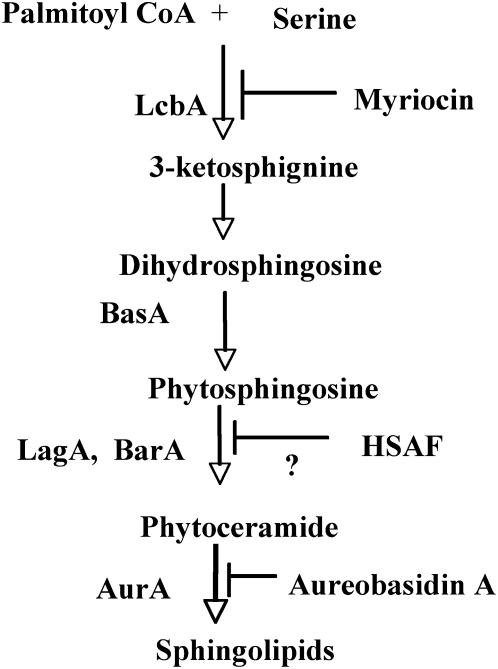
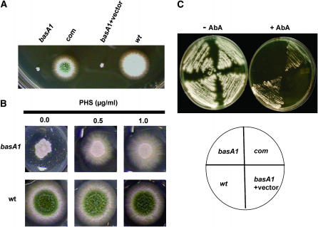
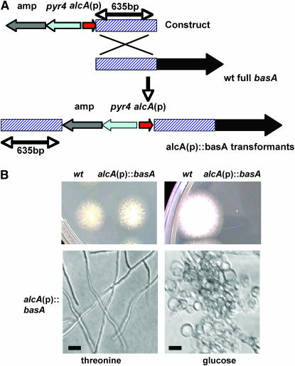
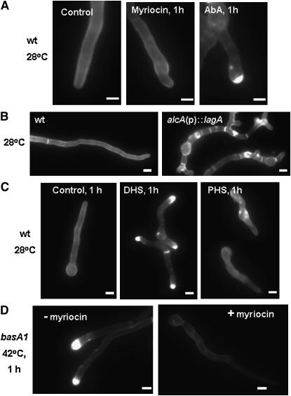
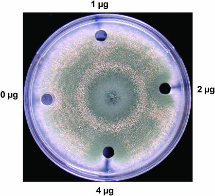
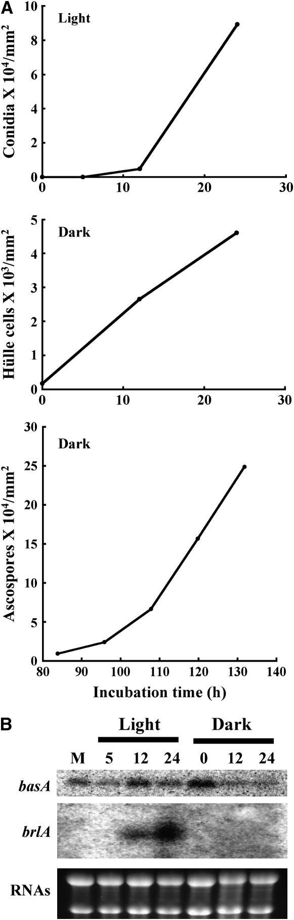
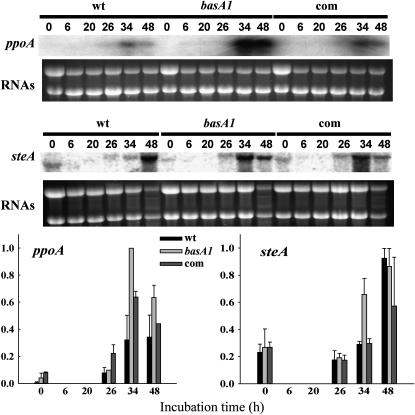
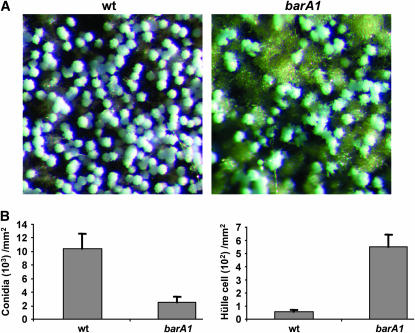
Sphingolipid C4 hydroxylase catalyzes the conversion of dihydrosphingosine to phytosphingosine. In Saccharomyces cerevisiae, Sur2 is essential for sphingolipid C4 hydroxylation activity but not essential for normal growth. Here we demonstrate that the Aspergillus nidulans Sur2 homolog BasA is also required for phytosphingosine biosynthesis but is also essential for viability. We previously reported that a point missense mutation in basA resulted in aberrant cell wall thickening. Here our data suggest that accumulation of dihydrosphingosine is responsible for this phenotype. In addition, two different mutations in basA consistently accelerated the transition from asexual development to sexual development compared to the wild-type strain. The phenotype could be suppressed by exogenous addition of phytosphingosine. Northern analysis suggests that faster sexual development in the basA mutant might be due to a higher transcription level of ppoA and steA, genes demonstrated to coordinate a balance between asexual and sexual development in A. nidulans. Consistent with these findings, mutations in the ceramide-synthase-encoding genes barA and lagA also caused faster transition from asexual to sexual development, supporting the involvement of sphingolipid metabolism in fungal morphogenesis.


Figures

F<sc>igure</sc> 1.—
Figure 1.—
A simplified scheme for the sphingolipid synthesis pathway in A. nidulans. Enzymes and inhibitors for the corresponding reaction steps are listed on the left and right, respectively.
F<sc>igure</sc> 2.—
Figure 2.—
Functional characterization of basA. (A) Complementation of basA1 mutant. basA1 mutant, a complemented strain cotransformed with a basA PCR product, and plasmid pRG3-AMA1, a basA1 strain transformed with vector pRG3-AMA1 and wild-type strain (GR5), were grown at 42° for 3 days. (B) Restoration of wild-type growth of basA1 mutant with exogenous phytosphingosine on solid medium. Wild-type strain A28 or basA1 mutant conidiophores were inoculated on MAG with PHS at the indicated dosages and incubated at 42° for 3 days. (C) Sensitivity of basA1 mutant to AbA. Strains were streaked onto MAG plates with or without 0.25 μg/ml AbA and incubated at 28° for 3 days.
F<sc>igure</sc> 3.—
Figure 3.—
Essential role of basA in growth. (A). Schematic of the generation of alcA(p)-controlled strains. (B) Phenotypic characterization of the alcA(p)∷basA strain. Wild-type strain A28 and alcA (p)∷basA strain ASL6 conidiospores were inoculated on alcA(p)-inducing medium MNVTF (threonine) or repressing medium MNV (glucose) and incubated at 28° for 3 days. Microscopic images were captured under a light microscope with a ×10 objective. Bar, 10 μm.
F<sc>igure</sc> 4.—
Figure 4.—
Effects of sphingolipid synthesis disruption on cell wall structure. (A and C) Wild-type strain A28 conidia were germinated at 28° for 12 hr and then shifted to a drug-free YGV (control) or to YGV supplemented with 0.25 μg/ml AbA, 20 μg/ml of myriocin, 1 μg/ml dihydrosphingosine, or 1 μg/ml phytosphingosine and incubated for an additional 1 hr. (B) Wild-type strain A28 and alcA(p)∷lagA strain ASL10 conidia were grown in alcA(p)-repressing medium YGV at 42° for 13 hr. (D) basA1 mutant conidiospores were germinated in YGV at 28° for 12 hr and shifted to fresh YGV with or without 20 μg/ml of myriocin for an additional 1 hr of incubation at 42°. Hyphae were fixed and stained with Calcofluor. Bar, 3 μm.
F<sc>igure</sc> 5.—
Figure 5.—
Comparison of asexual and sexual differentiation between wild type and the basA1 mutant. Conidiospores of A28 (wt), basA1 mutant, or complementation strain (com) were inoculated at the center of YGT plates and incubated at 28° in constant dark for 7 days. (A–C) Images of full colonies. (D–F) Images of growth in the area 3.5 cm away from the inoculation origin (circled area in A–C) under a dissecting microscope with a ×6 objective. Conidiophores (in G and I) and Hülle nursing cells (in H) are labeled with arrows, respectively. (G–I) Samples were collected, crushed, and fixed. Images in G–I were captured under a light microscope with a ×40 objective. (J) Quantitative comparison of conidiospore, Hülle cell, and ascospore production among wild type, basA1 mutant, and complementation strain. Quantitative comparison of ascospore production among wild-type, basA1 mutant, and complementation strain was done 10 days after inoculation. Samples were harvested from circled regions in A–C.
F<sc>igure</sc> 6.—
Figure 6.—
Supplementation of PHS suppresses sexual sporulation in the basA1 mutant. Conidiospores were inoculated at the center of the MAG plates, and wells (7-mm diameter) were made 3 cm away from the inoculation point. A total of 50 μl of YGV with the indicated doses of PHS was added to the wells.
F<sc>igure</sc> 7.—
Figure 7.—
Correlation of basA transcription with fungal development. Conidiospores from wild-type strain FGSC4 were germinated in YGV liquid medium at 28° for 18 hr. Mycelium was harvested and then transferred onto YGV solid medium. Asexual development was induced by exposing plates to constant light, and sexual development was induced by sealing plates with parafilm and incubating in the dark for 20 hr. Samples were taken at various time points during asexual development (5–24 hr after shifting to solid medium) and sexual development (0–24 hr after 20 hr sexual induction) for RNA analysis. (A) Quantitative analysis of conidia, Hülle cells, and ascospores after induction of asexual and sexual development, respectively. (B) Northern analysis of basA transcripts. rRNAs stained with ethidium bromide were shown as an indicator for equal loading of RNA.
F<sc>igure</sc> 8.—
Figure 8.—
Effects of the basA mutation on the transcription of developmental genes in A. nidulans. The wild-type strain A28, the basA1 mutant 8-145, and the complementation strain were cultured in shaken conditions in YGT liquid medium for 20 hr and then shifted onto solid YGT medium. At that time, plates were sealed with parafilm and incubated at 28° in the dark to induce sexual development for 20 hr. Total RNA corresponding to wild type and basA1 strains was isolated at the time of the shift (t = 0), at 6 and 20 hr (during sexual development induction), and at 26, 34, and 48 hr after the shift (6, 14, and 28 hr after sexual development induction, respectively). Transcriptional levels of ppoA and steA were examined by Northern analysis. rRNAs stained with ethidium bromide are shown to indicate RNA loading. The experiment was repeated twice with similar results (average values are shown). Relative transcript levels were quantified by densitometry using Scion Image Beta 4.0.2.
F<sc>igure</sc> 9.—
Figure 9.—
Role of BarA in sporulation. Conidiospores from wild-type strain A773 and the barA1 mutant UV13p were inoculated at the center of the MAG plates. Plates were sealed with parafilm and incubated at 28° in the dark for 7 days. (A) Image of the colony surface 1 cm away from the growth front. The image was captured with a Spot Insight Color camera on a Leica MZ75 dissecting microscope with a ×5 objective. (B) Quantitative comparison of conidial and Hülle cell production in the imaged area.

Similar articles

Cited by

References

    1. Adams, T. H., M. T. Boylan and W. E. Timberlake, 1988. brlA is necessary and sufficient to direct conidiophore development in Aspergillus nidulans. Cell 54: 353–362. - PubMed
    1. Adams, T. H., J. K. Wieser and J. H. Yu, 1998. Asexual sporulation in Aspergillus nidulans. Microbiol. Mol. Biol. Rev. 62: 35–54. - PMC - PubMed
    1. Bae, J. H., J. H. Sohn, C. S. Park, J. S. Rhee and E. S. Choi, 2004. Cloning and functional characterization of the SUR2/SYR2 gene encoding sphinganine hydroxylase in Pichia ciferrii. Yeast 21: 437–443. - PubMed
    1. Barz, W. P., and P. Walter, 1999. Two endoplasmic reticulum (ER) membrane proteins that facilitate ER-to-Golgi transport of glycosylphosphatidylinositol-anchored proteins. Mol. Biol. Cell 10: 1043–1059. - PMC - PubMed
    1. Beeler, T., D. Bacikova, K. Gable, L. Hopkins, C. Johnson et al., 1998. The Saccharomyces cerevisiae TSC10/YBR265w gene encoding 3-ketosphinganine reductase is identified in a screen for temperature-sensitive suppressors of the Ca2+-sensitive csg2Delta mutant. J. Biol. Chem. 273: 30688–30694. - PubMed

Publication types

MeSH terms