. 2007 Jun;81(12):6632-42.
doi: 10.1128/JVI.02730-06. Epub 2007 Apr 4.

Identification of a novel C-terminal cleavage of Crimean-Congo hemorrhagic fever virus PreGN that leads to generation of an NSM protein

Affiliations

Identification of a novel C-terminal cleavage of Crimean-Congo hemorrhagic fever virus PreGN that leads to generation of an NSM protein

Louis A Altamura et al. J Virol. 2007 Jun.

Abstract

The structural glycoproteins of Crimean-Congo hemorrhagic fever virus (CCHFV; genus Nairovirus, family Bunyaviridae) are derived through endoproteolytic cleavage of a 1,684-amino-acid M RNA segment-encoded polyprotein. This polyprotein is cotranslationally cleaved into the PreGN and PreGC precursors, which are then cleaved by SKI-1 and a SKI-1-like protease to generate the N termini of GN and GC, respectively. However, the resulting polypeptide defined by the N termini of GN and GC is predicted to be larger (58 kDa) than mature GN (37 kDa). By analogy to the topologically similar M segment-encoded polyproteins of viruses in the Orthobunyavirus genus, the C-terminal region of PreGN that contains four predicted transmembrane domains may also contain a nonstructural protein, NSM. To characterize potential PreGN C-terminal cleavage events, a panel of epitope-tagged PreGN truncation and internal deletion mutants was developed. These constructs allowed for the identification of a C-terminal endoproteolytic cleavage within, or very proximal to, the second predicted transmembrane domain following the GN ectodomain and the subsequent generation of a C-terminal fragment. Pulse-chase experiments showed that PreGN C-terminal cleavage occurred shortly after synthesis of the precursor and prior to generation of the GN glycoprotein. The resulting fragment trafficked to the Golgi compartment, the site of virus assembly. Development of an antiserum specific to the second cytoplasmic loop of PreGN allowed detection of cell-associated NSM proteins derived from transient expression of the complete CCHFV M segment and also in the context of virus infection.


Figures

FIG. 1.
FIG. 1.
Processing of the M segment-encoded polyprotein of CCHFV strain IbAr10200. A schematic of the CCHFV M segment-encoded polyprotein is shown, with known and suspected cleavage sites indicated. Signal peptidase is thought to generate the N terminus of the polyprotein and may also liberate PreGC as indicated (32). The mucin-GP38 domains are liberated by SKI-1 cleavage following the RRLL cleavage site to generate the N terminus of GN at amino acid 520 (32, 39). A second cleavage event, perhaps also mediated by SKI-1 or a similar protease, produces the N terminus of GC at residue 1041 following the sequence RKPL (32). Further cleavage by furin following the RSKR motif separates the mucin-like domain from GP38 (31). The region defined by the N termini of GN and GC encodes a 58-kDa polypeptide having four predicted transmembrane domains, indicated by black bars. Since mature GN is approximately 37 kDa, an additional C-terminal processing site may exist between GN and GC, leading to the generation of an NSM protein. The uncertain boundaries of this putative NSM protein are indicated by a dashed line. The cylinders labeled TM 1 to TM 4 represent the four predicted transmembrane helices between the ectodomains of GN and GC. Amino acid boundaries for each helix were predicted with TMHMM 2.0 (37).
FIG. 2.
FIG. 2.
PreGN is cleaved at its C terminus. (A) Schematic of CCHFV full-length M polyprotein and PreGN glycoprotein constructs. “M” and “961” refer to the constructs M[GCV5] and PreGNV5(961), respectively, that are described in the text. Each construct possessed a C-terminal V5-His6 epitope cassette. 293T/17 cells were transfected with the indicated constructs and then cell lysates were prepared at 18 to 22 h posttransfection. A plasmid expressing green fluorescent protein (GFP) was used as a negative control. The samples were separated by SDS-PAGE using 10% Bis-Tris NuPAGE gels and MOPS-based running buffer. Proteins in cell lysates were immunoblotted with an anti-GN ectodomain polyclonal antiserum (B) or a monoclonal antibody directed against the V5 epitope tag (C). The locations of PreGN, GN, and the C-terminal fragment are indicated.
FIG. 3.
FIG. 3.
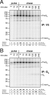
Pulse-chase analysis of PreGN cleavage events. 293T/17 cells were transfected with a plasmid expressing PreGNV5(961), metabolically labeled with [35S]cysteine-methionine for 15 min, and then placed in normal growth medium containing excess cysteine-methionine for the times indicated at the tops of the gels. The cells were then lysed and immunoprecipitated with anti-V5 epitope polyclonal serum (A) or an anti-GN ectodomain polyclonal serum (B). Some cells were labeled for only 5 or 10 min prior to lysis (left side of gel, as indicated). The samples were separated by SDS-PAGE using 10% Bis-Tris NuPAGE gels and MOPS-based running buffer. An asterisk indicates the location of the unidentified coprecipitating protein described in the text. The abundance of PreGN, GN, and the C-terminal fragment at each time point was determined by storage phosphor screen image analysis. Relative protein abundance represents the percentage of the maximum signal intensity obtained for each protein over the course of the experiment.
FIG. 4.
FIG. 4.
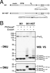
Trafficking and topology of the PreGN C-terminal fragment. (A) Schematic of PreGNV5 lumenal loop constructs. Using the PreGNV5(961) construct as a template, the lumenal loop was duplicated and an NST glycosylation sequon was added at the junction to make PreGNV5(961)-NST. Each construct possessed a C-terminal V5-His6 epitope cassette. (B) 293T/17 cells were transfected with the indicated constructs and cultured in the absence (top panel) or presence (bottom panel) of 1 μM DMJ, and then cell lysates were prepared at 18 to 22 h posttransfection. The samples were then treated with PNGase F, Endo H, or mock digested as indicated prior to separation by SDS-PAGE using 10% Bis-Tris NuPAGE gels and MOPS-based running buffer. Proteins in cell lysates were immunoblotted with a monoclonal antibody directed against the V5 epitope. The locations of PreGN, the C-terminal fragment of PreGNV5(961) (C-term961), and the deglycosylated (C-term961-NST) and glycosylated (C-term961-NSTglyc) C-terminal fragments of PreGNV5(961)-NST are indicated.
FIG. 5.
FIG. 5.
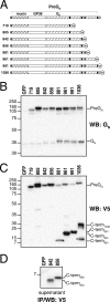
PreGN truncation mutants. (A) A schematic of the PreGNV5 truncation constructs is shown, with the predicted transmembrane domains indicated by black bars. The terminal amino acid for each construct (relative to the IbAr10200 M polyprotein) is indicated at the left, and each construct possessed a C-terminal V5-His6 epitope cassette. 293T/17 cells were transfected with the indicated constructs or with a plasmid expressing GFP, and then cell lysates were prepared at 18 to 22 h posttransfection. The samples were separated by SDS-PAGE using 10% Bis-Tris NuPAGE gels and MOPS-based running buffer. Proteins in the cell lysates were immunoblotted with an anti-GN ectodomain polyclonal antiserum (B) or a monoclonal antibody directed against the V5 epitope (C). An asterisk indicates a putative product of PreGN-PreGC cleavage in the PreGNV5(1036) construct. (D) For cells transfected with PreGNV5(842) and PreGNV5(856), proteins were also immunoprecipitated from the culture medium above these cells using a polyclonal antiserum directed against the V5 epitope. Afterwards, the precipitated proteins were separated by SDS-PAGE using 10% Bis-Tris NuPAGE gels and MES-based running buffer, followed by immunoblotting with a monoclonal antibody directed against the V5 epitope. The locations of PreGN and the C-terminal fragments (C-termxxx) are indicated (“xxx” refers to the terminal amino acid of the PreGNV5 truncation construct from which each fragment is derived).
FIG. 6.
FIG. 6.
PreGN internal deletion mutants. (A) A schematic of the PreGNV5 internal deletion constructs is shown, with the predicted transmembrane domains indicated by black bars. Using the PreGNV5(961) construct as a template, 18 to 22 amino acid deletions were made in cytoplasmic loops 1 and 2 as indicated. The lumenal loop was also deleted but then replaced with six glycines in order to maintain membrane topology of the protein. 293T/17 cells were transfected with the indicated constructs and then cell lysates were prepared at 18 to 22 h posttransfection. The samples were separated by SDS-PAGE using 10% Bis-Tris NuPAGE gels and MOPS-based running buffer. Proteins in the cell lysates were immunoblotted with anti-GN ectodomain polyclonal antiserum (B to D, top panels) or a monoclonal antibody directed against the V5 epitope (B to D, bottom panels). The locations of PreGN, GN, and the C-terminal fragment of PreGNV5(961) are indicated. GN and C-terminal fragment species having internal deletions are indicated by G and C-term961Δ, respectively.
FIG. 7.
FIG. 7.
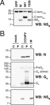
Identification of an NSM protein by transient expression and virus infection. (A) 293T/17 cells were transfected with pcDNA3.1 CCHFV glycoprotein expression constructs M[GCV5], PreGNV5(961), and PreGNV5(1036). The 30-kDa band present in all lanes is background, as it was also present in the GFP negative control. (B) 293T/17 cells were infected with CCHFV strain IbAr10200 at an MOI of 5, and cell lysates were prepared at approximately 24 h postinfection. In parallel, cells were also mock infected or transfected with a pCAGGS plasmid expressing the IbAr10200 M segment. Infected and mock culture supernatants were clarified and overlaid on a 20% sucrose-PBS cushion, and virions were semipurified by ultracentrifugation. C, cell lysate; P, virion pellet. The samples for panels A and B were separated by SDS-PAGE using 10% Bis-Tris NuPAGE gels and either MES-based (NSM) or MOPS-based (N, GN) running buffers. Proteins in cell and virion lysates were immunoblotted with anti-NSM cytoplasmic loop polyclonal antiserum (A and B, top panels), anti-N monoclonal antibody 9D5-1-1A (B, top panel), or anti-GN ectodomain polyclonal antiserum (B, middle panel).

References

    1. Ahmed, A. A., J. M. McFalls, C. Hoffmann, C. M. Filone, S. M. Stewart, J. Paragas, S. Khodjaev, D. Shermukhamedova, C. S. Schmaljohn, R. W. Doms, and A. Bertolotti-Ciarlet. 2005. Presence of broadly reactive and group-specific neutralizing epitopes on newly described isolates of Crimean-Congo hemorrhagic fever virus. J. Gen. Virol. 86:3327-3336. - PubMed
    1. Alexander, W. A., B. Moss, and T. R. Fuerst. 1992. Regulated expression of foreign genes in vaccinia virus under the control of bacteriophage T7 RNA polymerase and the Escherichia coli lac repressor. J. Virol. 66:2934-2942. - PMC - PubMed
    1. Bertolotti-Ciarlet, A., J. Smith, K. Strecker, J. Paragas, L. A. Altamura, J. M. McFalls, N. Frias-Staheli, A. Garcia-Sastre, C. S. Schmaljohn, and R. W. Doms. 2005. Cellular localization and antigenic characterization of Crimean-Congo hemorrhagic fever virus glycoproteins. J. Virol. 79:6152-6161. - PMC - PubMed
    1. Cash, P. 1985. Polypeptide synthesis of Dugbe virus, a member of the Nairovirus genus of the Bunyaviridae. J. Gen. Virol. 66:141-148. - PubMed
    1. Clerx, J. P., and D. H. Bishop. 1981. Qalyub virus, a member of the newly proposed Nairovirus genus (Bunyavividae). Virology 108:361-372. - PubMed

Publication types

LinkOut - more resources