. 2007 Jun;81(12):6207-15.
doi: 10.1128/JVI.00210-07. Epub 2007 Apr 4.

Base pairing between cis-acting sequences contributes to template switching during plus-strand DNA synthesis in human hepatitis B virus

Affiliations

Base pairing between cis-acting sequences contributes to template switching during plus-strand DNA synthesis in human hepatitis B virus

Eric B Lewellyn et al. J Virol. 2007 Jun.

Abstract

Hepadnaviruses utilize two template switches (primer translocation and circularization) during synthesis of plus-strand DNA to generate a relaxed-circular (RC) DNA genome. In duck hepatitis B virus (DHBV) three cis-acting sequences, 3E, M, and 5E, contribute to both template switches through base pairing, 3E with the 3' portion of M and 5E with the 5' portion of M. Human hepatitis B virus (HBV) also contains multiple cis-acting sequences that contribute to the accumulation of RC DNA, but the mechanisms through which these sequences contribute were previously unknown. Three of the HBV cis-acting sequences (h3E, hM, and h5E) occupy positions equivalent to those of the DHBV 3E, M, and 5E. We present evidence that h3E and hM contribute to the synthesis of RC DNA through base pairing during both primer translocation and circularization. Mutations that disrupt predicted base pairing inhibit both template switches while mutations that restore the predicted base pairing restore function. Therefore, the h3E-hM base pairing appears to be a conserved requirement for template switching during plus-strand DNA synthesis of HBV and DHBV. Also, we show that base pairing is not sufficient to explain the mechanism of h3E and hM, as mutating sequences adjacent to the base pairing regions inhibited both template switches. Finally, we did not identify predicted base pairing between h5E and the hM region, indicating a possible difference between HBV and DHBV. The significance of these similarities and differences between HBV and DHBV will be discussed.


Figures
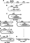
FIG. 1.
FIG. 1.
The reverse transcription pathway of HBV. (A) The pgRNA (thin black line) is the template for synthesis of minus-strand DNA. pgRNA is greater than genome length, so the termini are redundant (∼110 nt). DR1 and DR2 are 11-nt direct repeat sequences (open boxes), and there are two copies of DR1 due to its location in the terminal redundancy. The stem-loop, ɛ, is indicated. P protein forms a complex with ɛ, and the two are coencapsidated into a newly formed nucleocapsid. DNA synthesis takes place within the nucleocapsid. P protein acts as primer and polymerase to initiate the synthesis of minus-strand DNA. First, the first 3 (or 4) nt are synthesized using the bulge of ɛ as the template. (B) The nascent minus-strand DNA switches template to complementary sequence within DR1, and the synthesis of minus-strand DNA resumes. RNase H activity of P protein degrades the pgRNA as minus-strand DNA is synthesized. (C) The SS DNA intermediate contains a complete minus-strand DNA, and the 5′ portion of the pgRNA is not digested. This short RNA (∼17 nt) serves as the primer for the initiation of plus-strand DNA synthesis. (D) In the major pathway, the RNA primer switches template from DR1 to DR2 in a process termed primer translocation. Plus-strand DNA synthesis initiates from DR2. (E) When the nascent plus-strand DNA has extended to the 5′ end of the minus strand, it switches template from 5′r to 3′r in a process termed circularization. (F) The plus-strand DNA is extended from 3′r to subsequently form the mature RC DNA genome. (G) In the minor pathway, the RNA primer is used to initiate plus-strand DNA synthesis from DR1 in a process termed in situ priming. (H) The mature DL DNA genome results from fully elongated plus-strand DNA primed at DR1.
FIG. 2.
FIG. 2.
Locations of cis-acting sequences that contribute to the accumulation of RC DNA in HBV and DHBV relative to minus-strand DNA. (A) The locations of HBV cis-acting sequences that contribute to the accumulation of RC DNA are indicated (14, 16). Accumulation of RC DNA is less than 12% (black) or less than 75% (dark gray) of the WT reference level. Light gray indicates the previously understood boundaries of h3E and hM prior to this study. Previous mapping studies did not investigate the regions from nt 1845 to 1913 and 1745 to 1814 due to the presence of ɛ and other cis-acting sequences needed for minus-strand DNA synthesis (1, 2) (B) As a comparison, the locations of DHBV cis-acting sequences 3E, M, and 5E, which contribute to the accumulation of RC DNA (18). The M region can be further subdivided into M3 and M5, which make somewhat different contributions to the accumulation of RC DNA.
FIG. 3.
FIG. 3.
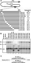
The hM region of HBV is localized to a short sequence. (A) Positions of deletion and substitution variants within the minus-strand DNA. The region from nt 2806 to 2994 is expanded to show relative sizes and positions of deletions and substitutions. The level of accumulation of RC DNA for each variant relative to the level of the WT reference and its standard deviation are shown on the right. Each variant was independently analyzed at least six times. NA, not available. (B) Representative Southern blot of hM region variants. RC, DL, and SS DNA forms are indicated. Asterisks indicate a new DNA form, seen only in some hM region variants, that is derived from a spliced RNA that has been encapsidated and reverse transcribed. The hybridization probe detected 350 nt of minus-strand DNA starting at nt 3096. (C) Calculation to determine the level of accumulation of RC DNA of each variant relative to the WT reference. The value “RC DNA” is the measurement of the DNA form labeled RC. The value “total minus-strand DNA” is the measurement of all DNA species between (and including) RC and SS DNA.
FIG. 4.
FIG. 4.
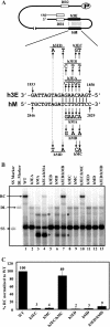
Base pairing between h3E and hM is necessary for the accumulation of RC DNA. (A) Predicted base pairing between h3E and hM. The substitution variants at either h3E or hM to test the contribution of the predicted base pairs are shown. (B) Southern blot analysis of substitution variants. Novel splice forms are indicated by asterisks. (C) Histogram of RC DNA accumulation efficiency for each variant. Error bars indicate standard deviations. Each variant was analyzed at least six times.
FIG. 5.
FIG. 5.
h3E and hM contribute to primer translocation and circularization. (A) Schematic representation of RC, DL, and SS DNA with relative positions of oligonucleotides used for primer extension analysis. (B to D) Primer extension gel with oligonucleotides 1, 2, and 3, respectively. The sequencing ladder is on the left. Bands representing replicative intermediate (R.I.) and internal standard (I.S.) DNA are indicated. Each replicative intermediate signal is normalized to the internal standard signal. The sizes of products for oligonucleotide 1, oligonucleotide 2, and oligonucleotide 3 extended on viral DNA are 155 nt, 175 nt, and 219 nt, respectively. Products for oligonucleotide 1, oligonucleotide 2, and oligonucleotide 3 extended on internal standard DNA are 224 nt, 217 nt, and 261 nt, respectively. (E and F) Graphical representations of primer translocation (P.T.) efficiency (E) and circularization efficiency (F) are shown. Each variant was analyzed at least six times. (G) Calculation to determine the relative level of primer translocation. We defined primer translocation as the proportion of replicative intermediates containing minus-strand DNA that had initiated plus-strand DNA from DR2 and elongated it up to at least the circularization point (about 217 nt from DR2). (H) Calculation to determine the relative level of circularization. We defined circularization as the fraction of plus-strand DNA having undergone primer translocation that also circularized and extended to nt 1859 (34 nt from the circularization point).
FIG. 6.
FIG. 6.
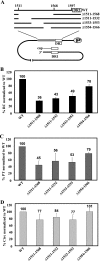
Efficiency of RC accumulation, primer translocation, and circularization for h5E region variants. (A) Positions of deletion and substitution variants within the minus-strand DNA. The region from nt 1511 to 1597 is expanded to show relative sizes and positions of deletions. (B to D) Histograms indicate the level of accumulation of RC DNA (B), primer translocation (PT) (C), and circularization (D) relative to the level of the WT reference. Error bars indicate standard deviations. Each variant was analyzed at least six times.

References

    1. Abraham, T. M., and D. D. Loeb. 2006. Base pairing between the 5′ half of ɛ and a cis-acting sequence, Φ, makes a contribution to the synthesis of minus-strand DNA for human hepatitis B virus. J. Virol. 80:4380-4387. - PMC - PubMed
    1. Bartenschlager, R., M. Junker-Niepmann, and H. Schaller. 1990. The P gene product of hepatitis B virus is required as a structural component for genomic RNA encapsidation. J. Virol. 64:5324-5332. - PMC - PubMed
    1. Bartenschlager, R., and H. Schaller. 1992. Hepadnaviral assembly is initiated by polymerase binding to the encapsidation signal in the viral RNA genome. EMBO J. 11:3413-3420. - PMC - PubMed
    1. Chen, Y., and P. L. Marion. 1996. Amino acids essential for RNase H activity of hepadnaviruses are also required for efficient elongation of minus-strand viral DNA. J. Virol. 70:6151-6156. - PMC - PubMed
    1. Cristofari, G., C. Bampi, M. Wilhelm, F. X. Wilhelm, and J. L. Darlix. 2002. A 5′-3′ long-range interaction in Ty1 RNA controls its reverse transcription and retrotransposition. EMBO J. 21:4368-4379. - PMC - PubMed

Publication types

LinkOut - more resources