. 2007 Jun;81(12):6573-83.
doi: 10.1128/JVI.02751-06. Epub 2007 Apr 4.

The Kaposi's sarcoma-associated herpesvirus K5 E3 ubiquitin ligase modulates targets by multiple molecular mechanisms

Affiliations

The Kaposi's sarcoma-associated herpesvirus K5 E3 ubiquitin ligase modulates targets by multiple molecular mechanisms

Robert E Means et al. J Virol. 2007 Jun.

Abstract

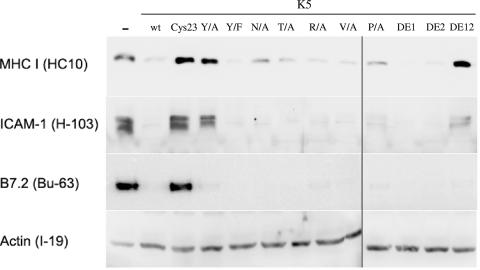
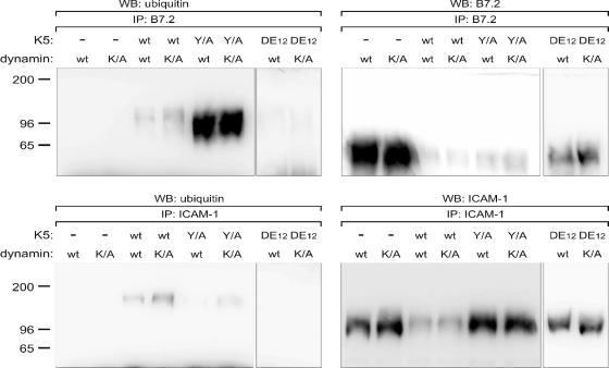
Kaposi's sarcoma-associated herpesvirus encodes two highly related membrane-associated, RING-CH-containing (MARCH) family E3 ubiquitin ligases, K3 and K5, that can down regulate a variety of cell surface proteins through enhancement of their endocytosis and degradation. In this report we present data that while K5 modulation of major histocompatibility complex class I (MHC-I) closely mirrors the mechanisms used by K3, alternative molecular pathways are utilized by this E3 ligase in the down regulation of intercellular adhesion molecule 1 (ICAM-1) and B7.2. Internalization assays demonstrate that down regulation of each target can occur through increased endocytosis from the cell surface. However, mutation of a conserved tyrosine-based endocytosis motif in K5 resulted in a protein lacking the ability to direct an increased rate of MHC-I or ICAM-1 internalization but still able to down regulate B7.2 in a ubiquitin-dependent but endocytosis-independent manner. Further, mutation of two acidic clusters abolished K5-mediated MHC-I degradation while only slightly decreasing ICAM-1 or B7.2 protein destruction. This same mutant abolished detectable ubiquitylation of all targets. These data indicate that while K5 can act as an E3 ubiquitin ligase to directly mediate cell surface molecule destruction, regulation of its targets occurs through multiple pathways, including ubiquitin-independent mechanisms.


Figures

FIG. 1.
FIG. 1.
K5 and mutant constructs. (A) The K5 protein is made up of three main modules as schematically represented in the center, with single-letter amino acid sequences shown for selected regions. At the N termini there is a C4HC3 RING-CH domain followed by two membrane-spanning domains (Tm). Carboxy terminal from the TMs is a CR. This conserved region is made up of a tyrosine-based motif (Tyr motif), a CM, a potential SH3B, and two stretches of acidic amino acids (DE1 and DE2). Numbering indicates the position within the K5 amino acid sequence, and letters above and below indicate residues that were mutated and the residue to which they were changed. Bars above or below the residues indicate mutant forms that contain mutations of multiple residues. (B) Expression of wild-type K5 proteins or each of the constructs was examined in stable BJAB cells. Normalized lysates were produced from each cell line and subjected to SDS-PAGE followed by Western blotting with an antibody against the V5 epitope tag encoded at the carboxy terminus of each construct.
FIG. 2.
FIG. 2.
Cell surface levels of MHC-I, ICAM-1, and B7.2 in stable K5-expressing BJAB cell lines. BJAB B cells stably expressing empty vector, wild-type K5, or K5 mutants, as indicated along the x axes, were stained with antibodies against MHC-I (A), ICAM-1 (B), or B7.2 (C) and analyzed by flow cytometry for mean channel fluorescence levels. Relative cell surface expression of each protein was determined by normalizing to the amount of fluorescence in empty vector cells. The data represent the averages of three separate experiments, with error bars indicating standard deviations.
FIG. 3.
FIG. 3.
Steady-state levels of target proteins in stable BJAB B-cell lines. Normalized lysates were prepared from BJAB B cells stably expressing empty vector, wild-type K5, or K5 mutants as indicated above each lane. For each cell line, 30 μg of total cell lysate was subjected to SDS-PAGE followed by Western blotting with the indicated antibody.
FIG. 4.
FIG. 4.
Pulse-chase analysis of target protein degradation. (A) Empty vector, K5 wild-type, K5 Cys23, K5 Y/A, and K5 DE12 cells were serum starved for 12 h followed by a 30-min labeling with [35S]methionine-cysteine. After washing with complete, cold medium, cells were transferred to 37°C for the indicated amounts of chase time before lysis in RIPA buffer. Lysates were then subjected to immunoprecipitation with antibodies against MHC-I (clone W6/32), ICAM-1 (H-103), or B7.2 (BU-63). After washing, precipitated proteins were subjected to SDS-PAGE followed by autoradiography using a Fuji LAS-1000 phosphorimager. Stars, lower-mobility products; M, mature form; P, precursor form. (B) Empty vector, K5 wild-type, K5 Cys23, K5 Y/A, and K5 DE12 cells were labeled and immunoprecipitated as described for panel A. After washing, the samples were heated to 100°C for 10 min in denaturation buffer, followed by addition of NP-40 to compete out excess SDS. The denatured proteins were then treated with 2,000 U PNGase F (New England Biolabs) for 1 h at 37°C followed by SDS-PAGE and autoradiography.
FIG. 5.
FIG. 5.
Quantitation of MHC-I degradation in BJAB B-cell lines. As described for Fig. 4, cells were labeled with [35S]methionine-cysteine and chased for the indicated amount of time, followed by immunoprecipitation with an anti-MHC-I antibody (clone W6/32). Precipitated proteins were subjected to SDS-PAGE and autoradiography followed by quantitation using a Fuji LAS-1000 phosphorimager. The amount of signal in each lane representing both the precursor and lower-mobility forms was normalized to the amount of signal in the time zero chase sample for each cell line. Data are representative of three separate experiments.
FIG. 6.
FIG. 6.
Quantitation of ICAM-1 and B7.2 degradation and export. Empty vector, K5 wild-type, K5 Cys23, K5 Y/A, K5 P/A, and K5 DE12 cells were treated as in described in the legend for Fig. 5; however, the lysates were immunoprecipitated with either an ICAM-1 (H-103)-specific (A) or B7.2 (BU-63)-specific (B) antibody. The total amount of signal corresponding to both the slowly (mature) and rapidly migrating (precursor) forms in each lane was normalized to the amount of signal for the time zero chase sample for each cell line. (C) The loss of the higher-mobility B7.2 immature form (labeled P) was quantitated from the same gels and normalized to the amount of precursor product at time zero. Data in each panel are representative of three separate experiments.
FIG. 7.
FIG. 7.
Endocytosis and down regulation of targets following electroporation of K5 or mutants. (A, B, and C) BJAB B cells were electroporated with constructs expressing GFP-fusion proteins of K5 wild-type, K5 Y/A, K5 DE12, or an unrelated viral protein, gH. At 90 min postelectroporation, cells were placed on ice and stained with unconjugated antibodies against MHC-I (A), ICAM-1 (B), or B7.2 (C) for 30 min. Cells were then transferred to 37°C at time zero, and samples were taken at the indicated time points. For each time point, cells were stained with the appropriate secondary antibody and subjected to flow cytometry. The resulting mean channel fluorescence (MCF) in the non-GFP-expressing population was used to normalize the MCF in the GFP-expressing population and displayed as the relative surface expression. (D, E, and F) In a parallel set of experiments, cells were electroporated with constructs expressing GFP-fusion proteins of K5 wild-type, K5 Y/A, or an unrelated viral protein, gH, and then incubated at 37°C for various amounts of time. At each time point cells were harvested onto ice, stained with conjugated antibodies against MHC-I (D), ICAM-1 (E), or B7.2 (F) for 30 min and then examined by flow cytometry. The resulting MCF in the non-GFP-expressing population was used to normalize the MCF in the GFP-expressing population and displayed as the relative surface expression. The data in each panel are representative of at least three separate experiments.
FIG. 8.
FIG. 8.
Ubiquitylation of B7.2, ICAM-1, and MHC-I in stable BJAB B-cell lines. Stable BJAB B-cell lines expressing empty vector, K5 wild-type, K5 Y/A, or K5 DE12 were electroporated with constructs expressing wild-type (wt) or dominant negative K44A mutant dynamin (K/A). At 48 h postelectroporation, live cells were purified on a Ficoll gradient and then subjected to immunoprecipitation with an antibody against B7.2 or ICAM-1, as indicated, followed by Western blotting for either ubiquitin (left panels) or the precipitated proteins (right panels).

Similar articles

Cited by

References

    1. Berger, S. L. 2002. Histone modifications in transcriptional regulation. Curr. Opin. Genet. Dev. 12:142-148. - PubMed
    1. Boname, J. M., and P. G. Stevenson. 2001. MHC class I ubiquitination by a viral PHD/LAP finger protein. Immunity 15:627-636. - PubMed
    1. Coscoy, L., and D. Ganem. 2000. Kaposi's sarcoma-associated herpesvirus encodes two proteins that block cell surface display of MHC class I chains by enhancing their endocytosis. Proc. Natl. Acad. Sci. USA 97:8051-8056. - PMC - PubMed
    1. Coscoy, L., and D. Ganem. 2001. A viral protein that selectively downregulates ICAM-1 and B7-2 and modulates T cell costimulation. J. Clin. Investig. 107:1599-1606. - PMC - PubMed
    1. Coscoy, L., D. J. Sanchez, and D. Ganem. 2001. A novel class of herpesvirus-encoded membrane-bound E3 ubiquitin ligases regulates endocytosis of proteins involved in immune recognition. J. Cell Biol. 155:1265-1273. - PMC - PubMed

Publication types