. 2007 Jun;81(12):6231-40.
doi: 10.1128/JVI.02859-06. Epub 2007 Apr 4.

Location and role of free cysteinyl residues in the Sindbis virus E1 and E2 glycoproteins

Affiliations

Location and role of free cysteinyl residues in the Sindbis virus E1 and E2 glycoproteins

Christopher B Whitehurst et al. J Virol. 2007 Jun.

Abstract

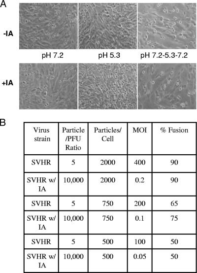
Sindbis virus is a single-stranded positive-sense RNA virus. It is composed of 240 copies of three structural proteins: E1, E2, and capsid. These proteins form a mature virus particle composed of two nested T=4 icosahedral shells. A complex network of disulfide bonds in the E1 and E2 glycoproteins is developed through a series of structural intermediates as virus maturation occurs (M. Mulvey and D. T. Brown, J. Virol. 68:805-812, 1994; M. Carleton et al., J. Virol. 71:1558-1566, 1997). To better understand the nature of this disulfide network, E1 and E2 cysteinyl residues were labeled with iodoacetamide in the native virus particle and analyzed by liquid chromatography-tandem mass spectrometry. This analysis identified cysteinyl residues of E1 and E2, which were found to be label accessible in the native virus particle, as well as those that were either label inaccessible or blocked by their involvement in disulfide bonds. Native virus particles alkylated with iodoacetamide demonstrated a 4-log decrease in viral infectivity. This suggests that the modification of free cysteinyl residues results in the loss of infectivity by destabilizing the virus particle or that a rearrangement of disulfide bonds, which is required for infectivity, is blocked by the modification. Although modification of these residues prevented infectivity, it did not alter the ability of virus to fuse cells after exposure to acidic pH; thus, modification of free cysteinyl residues biochemically separated the process of infection from the process of membrane fusion.


Figures

FIG. 1.
FIG. 1.
MS/MS identification of Cys-labeled peptide pairs obtained from differential alkylation of E1. The product ion spectra of several modified Cys peptides obtained by labeling native E1 at pH 8.0 using iodoacetamide (A and B) or denatured E1 using iodomethane (C and D) are shown. Each product ion spectrum (MS/MS spectrum) is a collection of ions produced by fragmentation of the intact peptide. Under the collision-induced dissociation conditions implemented in these measurements, fragmentation preferentially occurs along the backbone at peptide bonds to generate N-terminal fragments (b ions) and C-terminal fragments (y ions) at specific mass-to-charge (m/z) ratios and intensities which provide information regarding amino acid sequence and sites of modification. The predominant singly charged b and y product ion peaks are labeled accordingly, with the subscripts denoting their position within the identified peptide. Product ions eliciting neutral mass losses of H2O and NH3 are also indicated. Identified Cys residues are denoted as C* and C# for modifications corresponding to carboxamidomethylation and methylation, respectively. Analysis of the fragmentation patterns was performed by using SEQUEST with the corresponding scores presented in Table 1. The peptides were generated by in-gel proteolytic digestion of E1 using trypsin (A and C) or chymotrypsin (B and D) prior to extraction and LC/MS/MS analysis.
FIG. 2.
FIG. 2.
Representation of labeled cysteine residues in the amino acid sequence of the SV E1 and E2 proteins as determined by MS at pH 8.0. Boxed Cys residues were carboxamidomethylated by iodoacetamide. Underlined Cys residues were methylated by iodomethane. Boxed and underlined Cys residues represent alkylation by both reagents. RNase B served as a control.
FIG. 3.
FIG. 3.
Representation of labeled cysteine residues in the amino acid sequence of the SV E1 and E2 proteins as determined by S after a pH shift from 8.0 to 5.3 to 8.0. Boxed Cys residues were carboxamidomethylated by iodoacetamide. Underlined Cys residues were methylated by iodomethane. Boxed and underlined Cys residues represent alkylation by both reagents.
FIG. 4.
FIG. 4.
(A) Fusion of BHK cell monolayers by iodoacetamide-treated SV. In this example, 500 particles of iodoacetamide-treated (+IA) or untreated (−IA) SVHR/cell were placed on BHK monolayers and were examined for fusion at different pH values (see Materials and Methods). (B) Table relating the amounts of fusion observed at various concentrations of virus particles applied to BHK cells.

References

    1. Anthony, R. P., and D. T. Brown. 1991. Protein-protein interactions in an alphavirus membrane. J. Virol. 65:1187-1194. - PMC - PubMed
    1. Anthony, R. P., A. M. Paredes, and D. T. Brown. 1992. Disulfide bonds are essential for the stability of the Sindbis virus envelope. Virology 190:330-336. - PubMed
    1. Brown, D. T., and L. D. Condreay. 1986. Replication of alphaviruses in mosquito cells, p. 171-207. In M. J. Schlesinger (ed.), The Togaviridae and Flaviviridae. Plenum Publishing Corp., Inc., New York, NY.
    1. Carleton, M., and D. T. Brown. 1996. Disulfide bridge-mediated folding of Sindbis virus glycoproteins. J. Virol. 70:5541-5547. - PMC - PubMed
    1. Carleton, M., H. Lee, M. Mulvey, and D. T. Brown. 1997. Role of glycoprotein PE2 in formation and maturation of the Sindbis virus spike. J. Virol. 71:1558-1566. - PMC - PubMed

Publication types

LinkOut - more resources