Comparative Study
. 2007 Apr 4;27(14):3639-49.
doi: 10.1523/JNEUROSCI.4396-06.2007.

Beta-site amyloid precursor protein cleaving enzyme 1 levels become elevated in neurons around amyloid plaques: implications for Alzheimer's disease pathogenesis

Affiliations
Comparative Study

Beta-site amyloid precursor protein cleaving enzyme 1 levels become elevated in neurons around amyloid plaques: implications for Alzheimer's disease pathogenesis

Jie Zhao et al. J Neurosci. .

Abstract

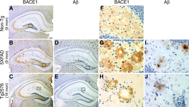
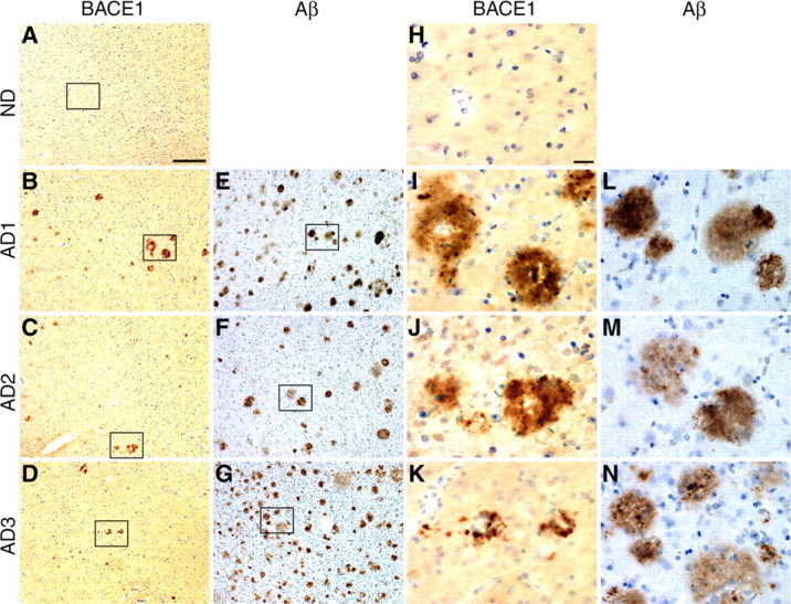
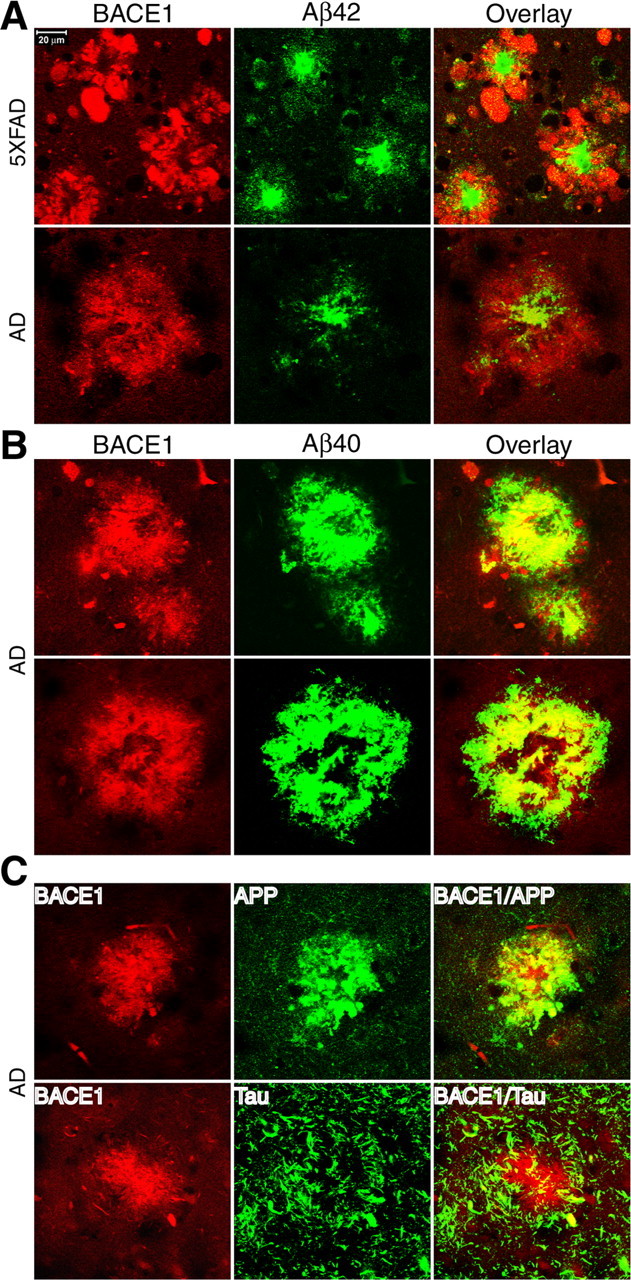
Beta-site amyloid precursor protein cleaving enzyme 1 (BACE1) (beta-secretase) initiates generation of beta-amyloid (Abeta), which plays an early role in Alzheimer's disease (AD). BACE1 levels are increased in postmortem AD brain, suggesting BACE1 elevation promotes Abeta production and AD. Alternatively, the BACE1 increase may be an epiphenomenon of late-stage AD. To distinguish between these possibilities, we analyzed BACE1 elevation using a highly specific BACE1 antibody, BACE-Cat1, made in BACE1-/- mice, which mount a robust anti-BACE1 immune response. Previous BACE1 immunohistochemical studies lack consistent results because typical BACE1 antibodies produce nonspecific background, but BACE-Cat1 immunolabels BACE1 only. BACE1 elevation was recapitulated in two amyloid precursor protein (APP) transgenic mouse lines. 5XFAD mice form amyloid plaques at young ages and exhibit neuron loss. In contrast, Tg2576 form plaques at a more advanced age and do not show cell death. These two mouse lines allow differentiation between early Abeta-induced events and late phenomena related to neuron death. BACE1 levels became elevated in parallel with amyloid burden in each APP transgenic, starting early in 5XFAD and late in Tg2576. The increase in BACE1 protein occurred without any change in BACE1 mRNA level, indicating a posttranscriptional mechanism. In APP transgenic and AD brains, high BACE1 levels were observed in an annulus around Abeta42-positive plaque cores and colocalized with neuronal proteins. These results demonstrate that amyloid plaques induce BACE1 in surrounding neurons at early stages of pathology before neuron death occurs. We conclude that BACE1 elevation is most likely triggered by the amyloid pathway and may drive a positive-feedback loop in AD.


Figures

Figure 1.
Figure 1.
Characterization of anti-BACE1 monoclonal antibodies generated in BACE1−/− mice. A, Schematic diagram of BACE1. BACE1 is a type I transmembrane glycoprotein with a signal sequence (SP) (residues 1–21) and a short prodomain (PD) (residues 22–45) that are cleaved during maturation, a long catalytic domain (residues 46–460), a single transmembrane domain (TM), and a short cytoplasmic tail (CD) (residues 478–501). The entire N-terminal catalytic domain (46–460) was expressed in E. coli and used as the antigen to immunize BACE1−/− mice and generate anti-BACE1 monoclonal antibodies. B, Screening and analysis of initial anti-BACE1 monoclonal antibodies in hybridoma culture supernatants by immunoblotting. Results for four representative hybridoma clones are shown. Protein extracts (15 μg/lane) of brain tissues from C57BL/6J wild-type (WT) mouse, BACE1−/− (KO) mouse, and non-demented aged human (Human) were immunoblotted and incubated with hybridoma culture media from clones 3D5, 2D5, 5C4, and 2D2. Compared with the others, supernatant staining by clone 3D5, subsequently named BACE-Cat1, displays the strongest BACE1 signal (arrow at left), has no nonspecific background bands in the BACE1 WT lane, and has no signal in the KO lane. Hybridoma supernatant dilutions are indicated in parentheses at the bottom of each blot. C, ELISA characterization of the BACE-Cat1 antibody. A dilution series of either affinity-purified BACE-Cat1 (1 mg/ml) or PA1-757 (1 mg/ml; ABR) antibodies were added to wells of a 96-well plate that were precoated with recombinant BACE1 catalytic domain (residues 46–460). Wells were then incubated with either HRP-conjugated goat anti-mouse or anti-rabbit secondary antibodies, developed, and optical densities (O. D.) read at 450 nm. Note the dose-dependent reduction in binding of BACE-Cat1 to BACE1 with increasing antibody dilutions, whereas PA1-757 (which recognizes the BACE1 C terminus) shows no detectable reading at any dilution and served as a negative control in the ELISA. D, The BACE-Cat1 antibody identifies authentic BACE1 immunoreactivity in mouse brain by immunohistochemistry. Coronal sections of C57BL/6J wild-type (WT) and BACE1−/− (KO; C57BL/6J background) brains were immunostained under identical conditions with BACE-Cat1 hybridoma culture supernatant and counterstained with hematoxylin after antigen retrieval (see Materials and Methods). In the WT brain section, diffuse BACE1 immunoreactivity is present throughout the entire section, but is higher in certain brain regions such as the mossy fiber pathway of the hippocampus (hatched arrow), the endopiriform nucleus/claustrum (open arrow), and globus pallidus/amygdala (closed arrow). In contrast, BACE-Cat1 signal does not appear in the KO brain section. Scale bar, 400 μm.
Figure 2.
Figure 2.
Immunoblotting with BACE-Cat1 reveals elevated BACE1 levels in human AD and APP transgenic mouse brains. A, Human autopsy brain tissues (temporal lobe cortex) of clinically diagnosed AD (n = 3) and non-demented controls (ND) (n = 3) were homogenized and analyzed by BACE-Cat1 immunoblot. Lanes with BACE1−/− (KO) brain homogenate and lysate from BACE1-overexpressing HEK293 cells (+) were included as negative and positive controls, respectively. Molecular mass markers are on the left. Immunoblot analysis for β-actin served as a loading control. B, Brain homogenates from representative 2- to 12-month-old 5XFAD (n = 3–5 per age) and age-matched normal control (Non-Tg) (n = 3–5 per age) mice were subjected to immunoblot analysis using BACE-Cat1 antibody. The BACE1 band migrates at ∼70 kDa, and the occasional band at ∼60 kDa represents endogenous mouse IgG (ms IgG) (detected by goat anti-mouse IgG secondary antibody) present in blood of brains from mice that were not transcardially perfused with buffer. Note that the lane with the homogenate from perfused KO brain does not have the background band. The intensities of the BACE1 bands in 5XFAD lanes are significantly greater than those of age-matched Non-Tg BACE1 bands. C, Quantification of 5XFAD and Non-Tg BACE1 signals detected by BACE-Cat1 immunoblot analysis is represented as percentage of 2 month Non-Tg control (100%). Note that BACE1 levels in 5XFAD brains reach ∼190% of control at 9 months and level off and that Non-Tg BACE1 levels appear to decrease slightly with age. Error bars indicate SEM. ***p < 0.001.
Figure 3.
Figure 3.
BACE-Cat1 reveals plaque-like staining in APP transgenic brains by immunohistochemistry. A–J, Coronal brain sections from 5XFAD and Tg2576 mice were immunostained with BACE-Cat1 antibody (A–C, F–H) or anti-total Aβ-amyloid antibody 4G8 (D, E, I, J) and counterstained with hematoxylin. Only images of the hippocampus are shown. A, F, Nontransgenic (Non-Tg) brain section from 9-month-old mouse stained with BACE-Cat1 shows the normal BACE1 immunoreactivity pattern with strongest labeling in the hippocampal mossy fiber pathway. B, G, 5XFAD brain section from 9-month-old mouse stained with BACE-Cat1 displays plaque-like BACE1 immunoreactivity. Note that BACE1-positive deposits typically have unstained cores. D, I, Adjacent 5XFAD brain section stained with 4G8 antibody reveals amyloid plaques that have a similar spatial distribution as BACE-Cat1-positive deposits. C, H, Tg2576 brain section from 18-month-old mouse stained with BACE-Cat1 also shows a plaque-like pattern of BACE1-positive deposits, although the number of deposits is less than in 5XFAD mice. E, J, Adjacent Tg2576 brain section immunostained with 4G8 antibody shows a similar distribution of amyloid plaques as that of BACE1-positive deposits. F–J, Higher magnification images of boxed areas in A–E. BACE1 immunoreactivity typically forms a ring-like structure with a clear center in both 5XFAD and Tg2576 transgenic mice (G, H), whereas amyloid plaques have a more uniformly stained core (I, J). Scale bars: A–E, 200 μm; F–J, 20 μm.
Figure 4.
Figure 4.
BACE-Cat1 reveals plaque-like staining in human AD brains by immunohistochemistry. A–N, Brain sections (inferior temporal gyrus or temporal lobe cortex) from normal (ND) and AD age-matched humans were immunostained with BACE-Cat1 (A–D, H–K) or anti-total Aβ antibody 4G8 (E–G, L–N) and counterstained with hematoxylin. A, H, Diffuse low-level BACE1 immunoreactivity with BACE-Cat1 is observed in ND cortex, but no deposits are apparent. B–D, Brain sections from three AD subjects all exhibit plaque-like BACE1-immunopositive deposits with BACE-Cat1, although numbers of BACE1 deposits in different patients vary considerably. I–K, As in APP transgenic brains, BACE1 immunoreactivity in AD brains is typically ring-like in appearance surrounding a relatively unstained core. E–G, AD brain sections stained with anti-total Aβ antibody 4G8 show numerous amyloid plaques in all three specimens. L–N, In contrast to BACE1-positive ring-like deposits, most amyloid plaques in AD brain exhibit stained cores with 4G8. Note that the number of BACE1-positive deposits is considerably less than that of amyloid plaques in the section of the same brain region and that many 4G8-positve deposits appear to be diffuse plaques. Higher-magnification images of boxed areas in A–G are shown in H–N. Scale bars: A–G, 60 μm; H–N, 20 μm.
Figure 5.
Figure 5.
Immunofluorescence double labeling of BACE1 together with Aβ, APP, or tau in 5XFAD and AD brains. Brain sections from 5XFAD or AD brains were costained with BACE-Cat1 and either antibodies against Aβ42 (A), Aβ40 (B), APP (C, top), or tau (C, bottom). A, 5XFAD (top) and AD (bottom) brain sections were double stained with BACE-Cat1 (red) and anti-Aβ42 C-terminal specific antibody (green). Note that BACE1 immunoreactivity surrounds the Aβ42 core of the plaques in both APP Tg and AD brains. B, AD brain section was double stained with BACE-Cat1 (red) and anti-Aβ40 C-terminal-specific antibody (green). Two representative amyloid plaques are shown. Note the high degree of colocalization between BACE1 and Aβ40 immunoreactivities. C, Top, AD brain section was double stained with BACE-Cat1 (red) and anti-APP C-terminal antibody (green). There is extensive colocalization of BACE1 and APP immunoreactivities within the deposit. C, Bottom, AD brain section was double stained with BACE-Cat1 (red) and anti-tau R1 antibody (green). Filamentous tau-positive structures tend to be concentrated around and partially overlap with the BACE1 deposit. Scale bar: A, Top left, 20 μm.
Figure 6.
Figure 6.
Immunofluorescence double labeling of BACE1 with neuronal and astrocytic markers in 5XFAD and AD brains. Brain sections from 5XFAD or AD brains were costained with BACE-Cat1 and either antibodies against NSE (A), synaptophysin (presynaptic marker) (B), MAP2 (postsynaptic marker) (C), β-III tubulin (neuronal marker) (D), or GFAP (astrocyte marker) (E). A, 5XFAD (top) and AD (bottom) brain sections were double stained with BACE-Cat1 (red) and anti-NSE antibody (green). Note that, within BACE1-positive deposits, BACE1 immunoreactivity colocalizes extensively (5XFAD) and moderately (AD) with NSE staining. B, 5XFAD (top) and AD (bottom) brain sections were double stained with BACE-Cat1 (red) and anti-synaptophysin antibody (green). Within deposits, BACE1 immunoreactivity colocalizes moderately well with synaptophysin staining in both 5XFAD and AD brains. C, 5XFAD brain sections were double stained with BACE-Cat1 (red) and anti-MAP2 antibody (green). BACE1 and MAP2 immunoreactivities do not colocalize to any significant extent. D, 5XFAD (top) and AD (bottom) brain sections were double stained with BACE-Cat1 (green) and anti-β-III tubulin antibody (red). BACE1 colocalization with β-III tubulin is present but relatively low within BACE1-positive deposits in 5XFAD and AD brains. E, 5XFAD (top) and AD (bottom) brain sections were double stained with BACE-Cat1 (red) and anti-GFAP antibody (green). Note that the overlap between BACE1 and GFAP immunoreactivities in 5XFAD and AD deposits is low. Scale bar: A, Top left, 20 μm.

References

    1. Apelt J, Bigl M, Wunderlich P, Schliebs R. Aging-related increase in oxidative stress correlates with developmental pattern of beta-secretase activity and beta-amyloid plaque formation in transgenic Tg2576 mice with Alzheimer-like pathology. Int J Dev Neurosci. 2004;22:475–484. - PubMed
    1. Binder LI, Frankfurter A, Rebhun LI. The distribution of tau in the mammalian central nervous system. J Cell Biol. 1985;101:1371–1378. - PMC - PubMed
    1. Blasko I, Beer R, Bigl M, Apelt J, Franz G, Rudzki D, Ransmayr G, Kampfl A, Schliebs R. Experimental traumatic brain injury in rats stimulates the expression, production and activity of Alzheimer's disease beta-secretase (BACE-1) J Neural Transm. 2004;111:523–536. - PubMed
    1. Bodendorf U, Danner S, Fischer F, Stefani M, Sturchler-Pierrat C, Wiederhold KH, Staufenbiel M, Paganetti P. Expression of human beta-secretase in the mouse brain increases the steady-state level of beta-amyloid. J Neurochem. 2002;80:799–806. - PubMed
    1. Cai H, Wang Y, McCarthy D, Wen H, Borchelt DR, Price DL, Wong PC. BACE1 is the major beta-secretase for generation of Abeta peptides by neurons. Nat Neurosci. 2001;4:233–234. - PubMed

Publication types

MeSH terms

Substances