Comparative Study
. 2007 Apr 4;27(14):3813-22.
doi: 10.1523/JNEUROSCI.5124-06.2007.

Dopamine receptor activation modulates GABA neuron migration from the basal forebrain to the cerebral cortex

Affiliations
Comparative Study

Dopamine receptor activation modulates GABA neuron migration from the basal forebrain to the cerebral cortex

James E Crandall et al. J Neurosci. .

Abstract

GABA neurons of the cerebral cortex and other telencephalic structures are produced in the basal forebrain and migrate to their final destinations during the embryonic period. The embryonic basal forebrain is enriched in dopamine and its receptors, creating a favorable environment for dopamine to influence GABA neuron migration. However, whether dopamine receptor activation can influence GABA neuron migration is not known. We show that dopamine D1 receptor activation promotes and D2 receptor activation decreases GABA neuron migration from the medial and caudal ganglionic eminences to the cerebral cortex in slice preparations of embryonic mouse forebrain. Slice preparations from D1 or D2 receptor knock-out mouse embryos confirm the findings. In addition, D1 receptor electroporation into cells of the basal forebrain and pharmacological activation of the receptor promote migration of the electroporated cells to the cerebral cortex. Analysis of GABA neuron numbers in the cerebral wall of the dopamine receptor knock-out mouse embryos further confirmed the effects of dopamine receptor activation on GABA neuron migration. Finally, dopamine receptor activation mobilizes striatal neuronal cytoskeleton in a manner consistent with the effects on neuronal migration. These data show that impairing the physiological balance between D1 and D2 receptors can alter GABA neuron migration from the basal forebrain to the cerebral cortex. The intimate relationship between dopamine and GABA neuron development revealed here may offer novel insights into developmental disorders such as schizophrenia, attention deficit or autism, and fetal cocaine exposure, all of which are associated with dopamine and GABA imbalance.


Figures

Figure 1.
Figure 1.
Neuronal migration assay using DiI labeling of migrating cells in forebrain slice preparations from E15 mice. A and B are bright-field images of representative rostral (A) and caudal (B) slices. DiI crystals (red clumps) were inserted approximately at the border between the lateral and medial ganglionic eminences (LGE and MGE, respectively) in rostral slices (A) and in the caudal ganglionic eminence (CGE) in caudal slices (B). In the rostral slices, the DiI-labeled cells originate in both the lateral and medial ganglionic eminences and migrate in multiple directions (red arrows), including toward the cerebral wall (CX). In the caudal slices, the DiI-labeled cells originate in the caudal ganglionic eminence and also migrate in multiple directions (red arrows), including toward the cerebral wall (CX). We counted the total number of DiI-labeled cells in the slices and calculated the percentage of cells entering the cerebral cortex (as a percentage of the total number of DiI-labeled cells in the slice). C–E are examples of slice preparations showing DiI-labeled cells (white arrows) that have migrated to the cerebral cortex (CX). The slices were exposed to medium without any drugs (control; C), medium containing the D1 receptor agonist SKF 81297 (1 μm; D), or the D2 receptor agonist quinpirole (20 μm; E) and maintained for 2 d in vitro. The DiI deposition site in the basal forebrain is visible (white asterisks). S, Septum; TH, thalamus, LV, lateral ventricle.
Figure 2.
Figure 2.
D1 receptor gain-of-function assay was performed by electroporating D1 receptor–EYFP constructs into the basal forebrain of E15 mouse brain slices. A, The location of the electroporation sites (green asterisks) is shown in diagrams of representative rostral and caudal slices. As in the DiI-labeling experiments (Fig. 1), the constructs were electroporated at the border between the lateral and medial ganglionic eminences (LGE and MGE, respectively) in rostral slices and in the caudal ganglionic eminence (CGE) in caudal slices. The electroporated cells scatter widely within 24 h, making it difficult to illustrate a prominent EYFP-positive electroporation site. Green arrows in A indicate that EYFP-labeled cells migrate in multiple directions, including toward the cerebral wall (CX). B–F, Examples of E15 mouse brain slices electroporated with the EYFP-labeled constructs and maintained in vitro for 2 d. A fluorescence image (B) is superimposed on the bright-field image (C) to register position of labeled cells with anatomical landmarks. Images in D–F are such superimposed images. Labeled cells are seen near the site of electroporation in the lateral ganglionic eminence (LGE; B–D), medial ganglionic eminence (D), caudal ganglionic eminence (E), and in the differentiating striatum (STR; white arrows in B and C; black arrows in D and E). Some electroporated, EGFP-positive cells have migrated to the cortex over the 2 d period (black arrows in E). The shaded areas in A represent the lateral ventricles. V, Lateral ventricle; HIP, hippocampus.
Figure 3.
Figure 3.
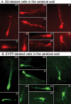
Morphology of migrating neurons. A, B, Cells with multiple morphological characteristics of migrating neurons were found entering the cerebral wall after DiI labeling (A) or electroporation of D1 receptor–EYFP constructs (B) in E15 forebrain slices. Examples are taken from different laminas of the cerebral wall of slices exposed to the D1 receptor agonist SKF 81297.
Figure 4.
Figure 4.
A–C, Examples of slice preparations from wild-type (WT; A), D1 receptor knock-out (D1−/−; B) and D2 receptor knock-out (D2−/−; C) E15 mouse embryos showing DiI-labeled cells (white arrows) that have migrated from the basal forebrain to the cerebral cortex (Cortex). The slices were exposed to dopamine (10 μm) plus ascorbic acid (0.01%) and maintained for 2 d in vitro. The DiI deposition site in the basal forebrain is visible (white asterisks). There are fewer DiI-labeled cells in the cortex of the D1 receptor knock-out mouse and relatively greater numbers in the cortex of the D2 receptor knock-out mouse, indicating the opposite effects of the loss of D1 or D2 receptor on neuronal migration.
Figure 5.
Figure 5.
GABA-positive cells in the cerebral wall of E15 mouse embryos. A–C, GABA immunohistochemistry was performed in coronal sections through the medial prefrontal cortex of littermate E15 wild type (WT; A); D1 receptor knock-out (D1−/−; B) and D2 receptor knock-out (D2−/−; C) mice. GABA-positive cells (black arrows) are distributed in all laminas of the cerebral wall. Overall, there are fewer GABA-positive profiles in the D1−/− cerebral wall (B) and greater numbers in the D2−/− cerebral wall (C) compared with the wild type (A). GABA labeling shows variability among the different laminas and across genotypes (especially SP). Quantitative comparisons (see Table 1) showed significant differences among the three genotypes in the IZ, VZ, and SVZ.
Figure 6.
Figure 6.
Dopamine receptor activation and neuronal cytoskeleton. Striatal neurons from E15 mice were grown in culture for 5 d and exposed to the D1 receptor agonist SKF 81297 or the D2 receptor agonist quinpirole. Immunocytochemistry was performed to analyze distribution of the motor protein CDHC or the cytoskeletal protein neuronal β-III tubulin (TuJ1). A, E, I, Nuclei were labeled with the DNA stain DAPI. B, C, In control cultures (B), CDHC was distributed throughout the cell body and the neurites (higher-magnification view in BA), and TuJ1 was principally localized to the neurites (C). D, Merged image of CDHC and TuJ1 labeling revealed overlap between the two proteins mainly in the neurites. F, In cultures exposed to SKF 81297, CDHC was distributed mainly to the soma, with prominent extensions into the proximal segments of the neurites near the cell soma (higher magnification in FA). G, In these cultures, TuJ1 was distributed to the neurites, as in control cultures. However, TuJ1 labeling was more intense in the neurites in the SKF 81297-exposed cultures compared with controls (C vs G). H, Merged images of the SKF 81297-exposed cultures revealed prominent tuft-like colocalization of CDHC and TuJ1, near the nucleus and extending into the proximal segments of the neurites (arrows in H). J, In cultures exposed to quinpirole, CDHC labeling was condensed around the nucleus (high-power view in jA). K, TuJ1 was distributed to neurites, as in control cultures (C), but, unlike in SKF 81297-exposed cultures, it was not localized to the cytoplasm. L, Merged images of CDHC and TuJ1 labeling did not reveal the prominent tuft-like colocalization. M, In summary, D1 receptor activation mobilizes CDHC and TuJ1 to produce a tuft-like pattern extending into the proximal segments of the neurites, whereas D2 receptor activation produces a condensation of the labeling around the nucleus. Scale bars: 15 μm.

Similar articles

Cited by

References

    1. Aizman O, Brismar H, Uhlen P, Zettergren E, Levey AI, Forssberg H, Greengard P, Aperia A. Anatomical and physiological evidence for D1 and D2 dopamine receptor colocalization in neostriatal neurons. Nat Neurosci. 2000;3:226–230. - PubMed
    1. Alifragis P, Liapi A, Parnavelas JG. Lhx6 regulates the migration of cortical interneurons from the ventral telencephalon but does not specify their GABA phenotype. J Neurosci. 2004;24:5643–5648. - PMC - PubMed
    1. Anderson SA, Eisenstat DD, Shi L, Rubenstein JLR. Interneuron migration from basal forebrain to neocortex: dependence on Dlx genes. Science. 1997;278:474–476. - PubMed
    1. Anderson SA, Marin O, Horn C, Jennings K, Rubenstein JL. Distinct cortical migrations from the medial and lateral ganglionic eminences. Development. 2001;128:353–363. - PubMed
    1. Ang ES, Jr, Haydar TF, Gluncic V, Rakic P. Four-dimensional migratory coordinates of GABAergic interneurons in the developing mouse cortex. J Neurosci. 2003;23:5805–5815. - PMC - PubMed

Publication types

MeSH terms