Comparative Study
. 2007 Apr 4;27(14):3921-32.
doi: 10.1523/JNEUROSCI.4710-06.2007.

Plexin-B2 controls the development of cerebellar granule cells

Affiliations
Comparative Study

Plexin-B2 controls the development of cerebellar granule cells

Roland H Friedel et al. J Neurosci. .

Abstract

Cerebellar granule cell progenitors proliferate postnatally in the upper part of the external granule cell layer (EGL) of the cerebellum. Postmitotic granule cells differentiate and migrate, tangentially in the EGL and then radially through the molecular and Purkinje cell layers. The molecular control of the transition between proliferation and differentiation in cerebellar granule cells is poorly understood. We show here that the transmembrane receptor Plexin-B2 is expressed by proliferating granule cell progenitors. To study Plexin-B2 function, we generated a targeted mutation of mouse Plexin-B2. Most Plexin-B2(-/-) mutants die at birth as a result of neural tube closure defects. Some mutants survive but their cerebellum cytoarchitecture is profoundly altered. This is correlated with a disorganization of the timing of granule cell proliferation and differentiation in the EGL. Many differentiated granule cells migrate inside the cerebellum and keep proliferating. These results reveal that Plexin-B2 controls the balance between proliferation and differentiation in granule cells.


Figures
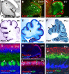
Figure 1.
Figure 1.
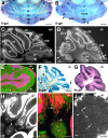
Plexin-B2 expression pattern in the developing cerebellum. All sections are sagittal. A, At E13, in situ hybridization with a DIG-labeled Plexin-B2 riboprobe showed that Plexin-B2 mRNA is highly expressed in the developing EGL (arrowheads) but is not expressed in the nuclear transitory zone (asterisk). B, E14 Plexin-B2+/− cerebellum immunostained for β-gal and RORα. β-gal is highly expressed in the EGL (arrowheads) above RORα-positive migrating Purkinje cells (asterisks). The weak staining in the ventricular zone is not specific. C, At E17, Plexin-B2 mRNA is still detected in the EGL (arrowheads) above CaBP-labeled Purkinje cells (asterisks). Plexin-B2 signal has been artificially colored in red using Photoshop (Adobe Systems, San Jose, CA) and superimposed to the CaBP labeling. D, At P0, Plexin-B2 transcripts are highly expressed in the EGL (arrowhead). E, β-gal expression is also confined to the EGL (arrowhead) in the cerebellum of P1 Plexin-B2+/−. F, In P7 Plexin-B2+/− cerebellum, β-gal expression is observed in both the EGL (arrowhead) and IGL (arrow). G, At P10, CaBP-positive Purkinje cells (arrowheads) form a monolayer. Plexin-B2 mRNA is only detected in the upper EGL (u) and not in the lower EGL (l). Plexin-B2 signal has been artificially colored in red using Photoshop and superimposed to the CaBP and Hoechst labeling. H, J, Wild-type cerebellum sections immunostained with anti-Plexin-B2 antibodies. H, At E15, Plexin-B2 is only detected in the EGL and choroid plexus (Hoechst counterstaining). I, P0 section labeled for Plexin-B2, H3P, and Hoechst. In the EGL, mitotic cells labeled with H3P (arrowheads) also express Plexin-B2. J, P10 section immunostained for Plexin-B2, CaBP, and TAG-1. Plexin-B2 is expressed in the upper EGL, whereas TAG-1 is found in the lower EGL and top of the upper part of the molecular layer above CaBP-positive Purkinje cells. Scale bars: A, 130 μm; B, 160 μm; C, 220 μm; D, 200 μm; E, 300 μm; F, 550 μm; G, 18 μm; H, 120 μm; I, 35 μm (I).
Figure 2.
Figure 2.
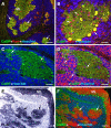
Cerebellar defects in Plexin-B2-deficient mice. Adult cerebella from Plexin-B2+/− mice (A, C) and Plexin-B2−/− mice (B, D–J). A, B, Cerebella were stained in toto for β-gal histochemistry. Vermis lobes are indicated by Roman numerals. In Plexin-B2−/−, the foliation is altered, some folia are fused (VI–VII), some fissures are absent, and others are less pronounced. In addition, ectopic β-gal-expressing cell clusters are found at the cerebellar surface (arrowhead). C, D, Sagittal cerebellum sections immunostained with anti-α6. In Plexin-B2−/−, lobules I–III and VI–VII are fused and the cerebellar lamination is profoundly perturbed. The precentral fissure (ce) and intercrural fissure (i) are absent. Islands of α6-unlabeled cells (arrows) are intermingled with α6-expressing granule cells. Ectopic clusters of α6-positive granule cells are found at the cerebellar surface (arrowheads). E, Higher magnification of a section double stained for α6 and CaBP. α6-positive granule cells are intermingled with CaBP-positive Purkinje cells. The arrowheads point to the superficial clusters of granule cells. F, β-gal staining also revealed the profound disorganization of the cerebellum and the islands of β-gal-negative cells. G, A section immediately adjacent to F was stained for PLAP. PLAP is expressed by parallel fibers in the molecular layer and in the β-gal-negative islands (arrowhead, compare with F). H, I, The base of some cerebellar folia is completely disorganized, and some dispersed α6-positive granule cells (arrowheads) invade the deep nuclei (asterisk) containing the CaBP-positive Purkinje cells axons. Some ectopic Purkinje cells are also found in the white matter (arrow in I). J, In the molecular layer of the less disorganized lobules, a few ectopic α6-positive granule cells (arrows) with two emerging dendrites (arrowheads) can be observed. Scale bars: A, B, 1.2 mm; C, D, 445 μm; E, 200 μm; F, G, 650 μm; H, 240 μm; I, 120 μm; J, 40 μm. S, Simplex; c1, crus1; c2, crus2; cu, culmen; pml, paramedial lobe; 1, primary fissure; py, prepyramidal fissure; 2, secondary fissure; po, posterior fissure; p, paraflocculus.
Figure 3.
Figure 3.
Cerebellum structure in Plexin-B2-deficient mice. Sagittal sections from adult Plexin-B2−/− mice. A, B, Sections immunostained for CaBP and parvalbumin and counterstained with Hoechst. A, Parvalbumin-labeled molecular layer interneurons are localized in the islands of ectopic CaBP-expressing Purkinje cells (arrows), dispersed among Hoechst-labeled granule cells. B, Higher magnification of a Purkinje cell island. Parvalbumin-positive interneurons are intermingled in Purkinje cell dendrites (arrowheads), and some typical basket cell “pinceaux” (arrow) are observed at the base of Purkinje cell bodies at the interface with granule cells. C, D, Section immunostained for CaBP (C) and vGlut2 (D) and counterstained with Hoechst. Ectopic CaBP-positive Purkinje cells (arrows in C) are contacted by vGlut2-labeled climbing fibers (arrows in D), whereas vGlut2-labeled mossy fiber rosettes (arrowheads in D) are found on granule cells. E, F, Section stained for PLAP histochemistry and immunostained for CaBP and counterstained with Hoechst. In F, PLAP staining was converted to red color using Photoshop. E, PLAP expression is concentrated in the molecular layer (ML, parallel fibers) and in the islands of ectopic Purkinje cells (arrows in E, F). Scale bars: A, 240 μm; B–D, 60 μm; E, F, 130 μm.
Figure 4.
Figure 4.
Radial glia, basal lamina, and granule cell migration in Plexin-B2-deficient mice. A–H, Sagittal cerebellum sections immunostained for GFAP (A–D, G, H), laminin (C–F), and Pax6 (G, H). Normal radial glia palisade in adult (A), P15 (C), or P11 (G) Plexin-B2+/−. In contrast, the radial glia palisade is very perturbed in adult (B), P15 (D), and P11 (H) Plexin-B2−/−, although radial glia fibers still extend to the pial surface and form typical end feet (arrowheads in D). C, D, Laminin expression at the GFAP-positive radial glia end feet is similar in P15 Plexin-B2+/− (C) and Plexin-B2−/− (D) mice. E, F, In adult, Plexin-B2+/− (E) and Plexin-B2−/− (F) laminin is highly expressed in the basal lamina covering each cerebellar folia. The staining is similar in heterozygous and homozygous. G, H, At P11, radially migrating Pax6-expressing granule cells can be observed apposed to GFAP-positive radial glia fibers both in Plexin-B2+/− and in Plexin-B2−/− (arrowheads in G, H). Scale bars: A, B, 270 μm; C, D, 50 μm; E, F, 220 μm; G, H, 35 μm.
Figure 5.
Figure 5.
Development of cerebellar defects in Plexin-B2-deficient mice. Sagittal sections of the cerebellum of Plexin-B2+/− (A, C, E, G, I–L, Q, R) and Plexin-B2−/− (B, D, F, H, M–P, S, T) mice. A, B, Section of E14 embryos immunostained with Pax6 and β-gal antibodies. The development of the EGL (arrowheads) and the distribution of granule cell progenitors coexpressing Pax6 and β-gal is similar in Plexin-B2+/− and Plexin-B2−/− mice. C, D, Higher magnification of the EGL of E14 cerebellum double labeled with β-gal and RORα antibodies. In both Plexin-B2+/− (C) and Plexin-B2−/− (D) cerebella, β-gal-expressing granule cell progenitors are restricted to the EGL above RORα-positive Purkinje cells. E, F, At E17, Pax6 expression pattern in the cerebellum is comparable in Plexin-B2+/− (E) and Plexin-B2−/− (F). G, H, GFP expression in whole-mount cerebella from E17 Math1:GFP;Plexin-B2+/− (G) and Math1:GFP;Plexin-B2−/− (H) mice. A fissure (arrow) is lacking on the rostral cerebellum of Plexin-B2−/− mice. I–P, P0 Plexin-B2+/− (I–L) or Plexin-B2−/− (M–P) cerebellum sections hybridized with Math1 riboprobe (I, M) or immunostained with Sema6A (J, L, N, P) and TAG-1 (K, L, O, P) antibodies. I, Math1 mRNA is only expressed in granule cell progenitors in the EGL (arrowheads). J–L, Sema6A and TAG-1 are expressed in postmitotic granule cells in the lower EGL (l in L) and not in the upper EGL (u in L). However, although Sema6A is expressed in a decreasing anteroposterior gradient, TAG-1 is homogeneously expressed in the EGL. M, In Plexin-B2−/− cerebellum, Math1 mRNA expression is still confined to the EGL that has a normal anteroposterior distribution (arrowheads). N–P, Likewise, Sema6A and TAG-1 are still coexpressed, and Sema6A expression respects its normal gradient. However, Sema6/TAG-1-immunostained cells are also observed outside the EGL in the cerebellar parenchyma (arrowheads in N, O). In addition, the upper and lower EGL are mixed, and streams of TAG-1/Sema6A-expressing cells invade the cerebellum (arrowheads in P). Q, R, Section of P0 Plexin-B2+/− cerebellum stained for PLAP and immunolabeled for CaBP (R). In R, PLAP staining was converted to red color with Photoshop. PLAP-expressing cells are restricted to the EGL (arrowheads in Q) and never observed in CaBP-positive Purkinje cells (arrowheads in R). S, T, In P0 Plexin-B2−/−, the foliation is less pronounced. Most PLAP-expressing cells are in the EGL (arrowhead in S), but many are also detected within the cerebellar parenchyma (arrow in S). T, The ectopic PLAP-expressing granule cells (arrow) are intermingled with CaBP-positive Purkinje cell bodies (arrowhead; inset in S). Scale bars: A, B, E, F, 175 μm; C, D, 150 μm; G, H, 880 μm; I–K, M–O, Q, S, 300 μm; L, P, 45 μm; R, T, 80 μm.
Figure 6.
Figure 6.
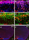
Cell proliferation in neonatal Plexin-B2-deficient mice. Sagittal sections from P0 Plexin-B2+/− (A, C, E) and Plexin-B2−/− (B, D, F) mice immunostained for Ki67 (A, B), Pax6 and BrdU (C, D), or H3P (E, F) and counterstained with Hoechst (A, B, E, F). A, C, In Plexin-B2+/− mice, proliferating granule cell progenitors labeled with Ki67 and BrdU are mostly found in the upper EGL (u), whereas postmitotic granule cells (only Pax6 and Hoechst labeled) start to appear in the lower EGL (l). Some proliferating cells (Pax6 negative) are also found outside the EGL and most likely correspond to precursors for molecular interneurons and oligodendrocytes (B, D). In contrast, in Plexin-B2−/− animals, the upper and lower EGL are not segregated, and proliferating Pax6-positive granule cells are also observed away from the EGL deeper inside the cerebellar cortex. E, F, H3P immunostaining in P0 cerebellum. Scale bars: A–D, 17 μm; E, F, 120 μm.
Figure 7.
Figure 7.
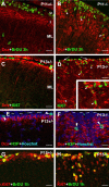
EGL defects in postnatal Plexin-B2-deficient mice. All sections are sagittal. A–D, P11 (A, B) or P13 (C–F) cerebellum sections of Plexin-B2+/− (A, C, E) and Plexin-B2−/− (B, D, F) mice immunostained for Dcx and BrdU (injected 3 h before fixation; A, B) or Ki67 (C, D) or H3P (E, F). In the EGL of Plexin-B2+/−, BrdU-labeled cells and Ki67- or H3P-immunoreactive cells are only found in the upper EGL (u; arrowhead in E), whereas Dcx is expressed in the lower EGL (l) and molecular layer (ML). In contrast, in Plexin-B2−/−, BrdU-, H3P-, or Ki67-labeled cells are also found in the molecular layer (arrowheads in B, D, F), whereas Dcx-immunoreactive cells are observed near the pial surface (arrows in D). Note also the reduction of size of the EGL between P11 and P13 in Plexin-B2+/−. G, H, P15 cerebellum sections of Plexin-B2+/− (G) and Plexin-B2−/− (H) mice immunostained for Ki67 and BrdU (injected 1 h before fixation). In Plexin-B2−/−, many double-labeled cells are observed away from the pial surface. Scale bars: A–D, 25 μm; E–H, 15 μm.
Figure 8.
Figure 8.
Ectopic granule cell proliferation in Plexin-B2-deficient mice. All sections are sagittal. A, B, E, P13 Plexin-B2−/− cerebellum immunostained for Ki67 and Hoechst (B) and Pax6 (E). Ki67-labeled proliferating cells are found in the EGL (arrows in A, B) but also in the periphery of Purkinje cell islands (asterisks) at the interface with granule cells (arrowheads). Most Ki67-positive cells are also labeled for Pax6 (arrowheads in E). C, D, P11 Plexin-B2+/− (C) and Plexin-B2−/− (D) cerebella hybridized with a Math1 riboprobe. Math1 expression is only detected in the EGL (arrows). In Plexin-B2−/−, gaps in Math1 expression are also observed (arrowheads in D). F–H, P13 Plexin-B2+/− (F) and Plexin-B2−/− (G, H) cerebellum hybridized with Barhl1 riboprobe. In H, Barhl1 signal has been converted to red color with Photoshop and superposed to CaBP immunostaining and Hoechst staining. In Plexin-B2+/−, Barlh1 mRNA is highly expressed in granule cell progenitors in the EGL (arrow) and at a low level in the molecular layer (ML) and IGL. Barhl1 expression is also very high in the EGL of Plexin-B2−/− (arrow) and at a low level the IGL and at the periphery (arrowheads) of the ectopic islands of CaBP-positive Purkinje cells (asterisks). I–K, Quantifications of the number of proliferating cells at P11 (I) or P15 (J, K) revealed by short-term BrdU pulse labeling (I, J; see Materials and Methods) or H3P immunostaining (K). Number of cells are expressed by millimeters of sections (J, K) or by 60× microscope field (I; see Materials and Methods). Statistics are based on n = 2 animals for each cases. Scale bars: A, 150 μm; B, 60 μm; C, D, 30 μm; E, 20 μm; F–H, 40 μm.
Figure 9.
Figure 9.
Model of Plexin-B2 function in the developing cerebellum. A, In newborn wild-type mice, granule cell progenitors (in red) proliferate exclusively in the upper EGL (U). Postmitotic granule cells start differentiating and migrate in the lower EGL (L) before migrating radially through the molecular layer (ML) and the Purkinje cell layer (PCL) to the IGL. At this stage, Purkinje cells (in gray) are still distributed in multiple layers. B, In newborn Plexin-B2 mutants, cells of the upper and lower EGL are intermingled, and cells expressing markers of differentiated granule cells divide during their migration in the molecular layer and also inside the Purkinje cell layers. C, In adult mice, Purkinje cells are aligned in a monolayer, above differentiated granule cell bodies (in blue), all localized in the IGL. D, In adult Plexin-B2 mutants, some ectopic granule cells are found at the top of the molecular layer and the cerebellar cortex is fragment. Islands of Purkinje cells (arrow) are embedded in differentiated granule cells.

Similar articles

Cited by

References

    1. Alder J, Lee KJ, Jessell TM, Hatten ME. Generation of cerebellar granule neurons in vivo by transplantation of BMP-treated neural progenitor cells. Nat Neurosci. 1999;2:535–540. - PubMed
    1. Artigiani S, Conrotto P, Fazzari P, Gilestro GF, Barberis D, Giordano S, Comoglio PM, Tamagnone L. Plexin-B3 is a functional receptor for semaphorin 5A. EMBO Rep. 2004;5:710–714. - PMC - PubMed
    1. Aruga J, Inoue T, Hoshino J, Mikoshiba K. Zic2 controls cerebellar development in cooperation with Zic1. J Neurosci. 2002;22:218–225. - PMC - PubMed
    1. Aurandt J, Vikis HG, Gutkind JS, Ahn N, Guan KL. The semaphorin receptor plexin-B1 signals through a direct interaction with the Rho-specific nucleotide exchange factor, LARG. Proc Natl Acad Sci USA. 2002;99:12085–12090. - PMC - PubMed
    1. Blaess S, Corrales JD, Joyner AL. Sonic hedgehog regulates Gli activator and repressor functions with spatial and temporal precision in the mid/hindbrain region. Development. 2006;133:1799–1809. - PubMed

Publication types

LinkOut - more resources