. 2007 Jun;18(6):2244-53.
doi: 10.1091/mbc.e06-11-0998. Epub 2007 Apr 4.

Active Arf6 recruits ARNO/cytohesin GEFs to the PM by binding their PH domains

Affiliations

Active Arf6 recruits ARNO/cytohesin GEFs to the PM by binding their PH domains

Lee Ann Cohen et al. Mol Biol Cell. 2007 Jun.

Abstract

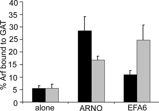
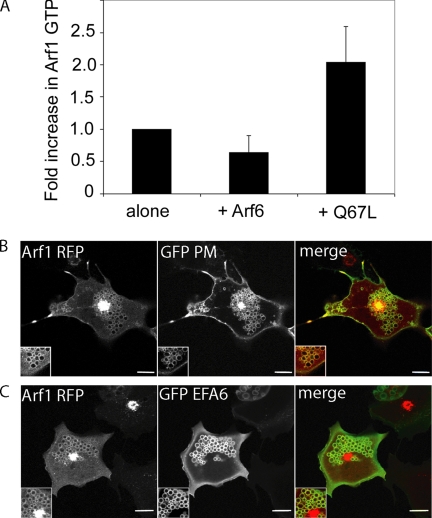
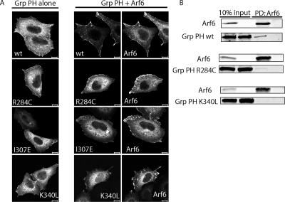
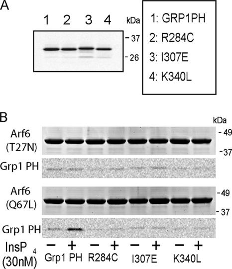
ARNO is a soluble guanine nucleotide exchange factor (GEF) for the Arf family of GTPases. Although in biochemical assays ARNO prefers Arf1 over Arf6 as a substrate, its localization in cells at the plasma membrane (PM) suggests an interaction with Arf6. In this study, we found that ARNO activated Arf1 in HeLa and COS-7 cells resulting in the recruitment of Arf1 on to dynamic PM ruffles. By contrast, Arf6 was activated less by ARNO than EFA6, a canonical Arf6 GEF. Remarkably, Arf6 in its GTP-bound form recruited ARNO to the PM and the two proteins could be immunoprecipitated. ARNO binding to Arf6 was not mediated through the catalytic Sec7 domain, but via the pleckstrin homology (PH) domain. Active Arf6 also bound the PH domain of Grp1, another ARNO family member. This interaction was direct and required both inositol phospholipids and GTP. We propose a model of sequential Arf activation at the PM whereby Arf6-GTP recruits ARNO family GEFs for further activation of other Arf isoforms.


Figures

Figure 1.
Figure 1.
Activation of Arf1 and Arf6 by ARNO and EFA6 in cells. Arf1-HA or Arf6-HA were expressed in HeLa cells either alone or in the presence of Flag-ARNO or Flag-EFA6. The fraction of Arf1 (■) or Arf6 (▩) that was GTP-bound was determined using a GGA3-GAT pulldown assay as described in Materials and Methods. Shown are the means and SD of triplicate experiments.
Figure 2.
Figure 2.
ARNO recruits Arf1 to the PM to form ruffles and macropinosomes. (A) HeLa cells expressing Arf1-RFP or GFP-ARNO were expressed by themselves or together and imaged live 24 h after transfection. (B) COS-7 cells coexpressing Arf1-RFP and GFP-ARNO (3G) were imaged at 15-s intervals for 30 min at 37°C. (C) COS-7 cells expressing Arf1-RFP and GFP-EFA6 were imaged as above. Shown in B and C are images from a selected 150-s interval from Supplementary Videos 1 and 2, respectively. Bars, 10 μm.
Figure 3.
Figure 3.
ARNO is recruited to the PM by GTP-bound Arf6. (A) HeLa cells expressing Flag-ARNO alone, or coexpressing Flag-ARNO and either Arf6, Arf6T27N, or Arf6Q67L, were processed for immunofluorescence as described and labeled with antibodies to Flag and Arf6. (B) Arf1-RFP, GFP-ARNO, and Arf6 were all expressed in HeLa cells. Twenty-four hours after transfection, cells were imaged live for ARNO and Arf1, which both localized to vesicle structures that form with overexpression of Arf6. (C) Flag-GRP1 was expressed alone or with Arf6Q67L and cells were immunolabeled with antibodies to Flag and Arf6. (D) Flag-ARNO was expressed with myc-PIP 5-kinase Iα, and cells were immunolabeled with antibodies to Flag and myc. (E) GFP-ARNO was expressed alone or with HA-tagged Arf6, Arf6T27N, or Arf6Q67L. Lysates from these cells were probed with anti-HA agarose to precipitate the HA-tagged Arfs, and the pulldowns were examined by Western blot against HA (Arf6) and GFP (ARNO) as described in Materials and Methods. Bars, 10 μm.
Figure 4.
Figure 4.
Active Arf6 leads to activation of Arf1. (A) Arf1-HA was expressed in HeLa cells either alone or in the presence of Arf6 or Arf6Q67L. The fraction of Arf1 that was GTP-bound was determined using a GGA3-GAT pulldown assay. Shown is the average fold-increase in Arf1-GTP from four independent experiments ±1 SD. (B) Arf1-RFP, Arf6Q67L, and GFP-PM were coexpressed in Cos-7 cells, and cells were imaged live 24 h after transfection. (C) Arf1-RFP, GFP-EFA6, and Arf6 were coexpressed in Cos-7 cells and imaged 18 h after transfection. Bars, 10 μm.
Figure 5.
Figure 5.
The PH domain of ARNO interacts with GTP-bound Arf6. (A) A flag-tagged catalytically inactive mutant of ARNO (ARNO EK), ARNO lacking the PH domain (ARNO ΔPH), GFP-tagged PH domain of ARNO triglycine variant (PH(3G)), PH domain of ARNO diglycine variant (PH(2G)) or GFP-tagged PH domain of Btk (Btk PH) were expressed alone or coexpressed with Arf6 in HeLa cells and stained with rabbit anti-Arf6 and either anti-Flag antibody (for ARNO EK and ARNO ΔPH) or mouse anti-GFP (for ARNO PH domains and Btk PH domain). For quantification of recruitment onto membranes see Supplementary Figure 1. (B) GFP-ARNO PH(3G) was expressed either alone or coexpressed with wt Arf1-HA or its mutants, or wt Arf6-HA or its mutants. Lysates from these cells were probed with anti-HA agarose, and immunoprecipitations were examined by Western blot using antibodies to HA (Arfs) and GFP (ARNO PH(3G)). (C) GFP-ARNO PH(2G) was expressed either alone or coexpressed with wt Arf6-HA or its mutants and examined with an anti-HA agarose pulldown as described. Bars, 10 μm.
Figure 6.
Figure 6.
Recruitment of the PH domain of Grp1 to the PM requires an interaction with Arf6 and the ability to bind phospholipids. (A) YFP-Grp1 PH domain or its mutants were expressed alone or coexpressed with Arf6 in HeLa cells and stained with antibodies to Arf6 and GFP (Grp1 PH domains). For quantification of recruitment onto membranes see Supplementary Figure 1. (B) YFP-Grp1 PH domain or its mutants were expressed alone or coexpressed with Arf6-HA in HeLa cells. Lysates from these cells were probed with anti-HA agarose to precipitate Arf6-HA, and pulldowns were examined by Western blot with antibodies to HA (Arf6) and GFP (Grp1 PH domain). Bars, 10 μm.
Figure 7.
Figure 7.
The interaction between Arf6-GTP and the Grp PH domain is direct and requires phosphoinositide. (A) Fluorescence scan of 20 μg purified YFP Grp1 PH domain or its mutants R284C, I307E, or K340L (B) Twenty micrograms of purified YFP Grp1 PH domain or its mutants was incubated with 20 μg purified GST-Arf6 T27N or GST-Arf6 Q67L immobilized on glutathione Sepharose beads in the presence (+) or absence of 30 nM Ins(1,3,4,5)P4. Bound proteins were resolved by SDS-PAGE, scanned for fluorescence of the YFP PH domains, and subsequently stained with Coomassie to visualize the GST-Arf6. The wt YFP Grp1 PH domain bound to GST-Arf6Q67L only in the presence of Ins (1,3,4,5)P4.

Similar articles

Cited by

References

    1. Antonny B., Beraud-Dufour S., Chardin P., Chabre M. N-terminal hydrophobic residues of the G-protein ADP-ribosylation factor-1 insert into membrane phospholipids upon GDP to GTP exchange. Biochemistry. 1997;36:4675–4684. - PubMed
    1. Balla T. Inositol-lipid binding motifs: signal integrators through protein-lipid and protein-protein interactions. J. Cell Sci. 2005;118:2093–2104. - PubMed
    1. Beemiller P., Hoppe A. D., Swanson J. A. A phosphatidylinositol- 3-kinase-dependent signal transition regulates ARF1 and ARF6 during Fcgamma receptor-mediated phagocytosis. PLoS Biol. 2006;4:e162. - PMC - PubMed
    1. Beraud-Dufour S., Robineau S., Chardin P., Paris S., Chabre M., Cherfils J., Antonny B. A glutamic finger in the guanine nucleotide exchange factor ARNO displaces Mg2+ and the beta-phosphate to destabilize GDP on ARF1. EMBO J. 1998;17:3651–3659. - PMC - PubMed
    1. Brown F. D., Rozelle A. L., Yin H. L., Balla T., Donaldson J. G. Phosphatidylinositol 4,5-bisphosphate and Arf6-regulated membrane traffic. J. Cell Biol. 2001;154:1007–1017. - PMC - PubMed

Publication types

MeSH terms