. 2007 Apr 26;446(7139):1091-5.
doi: 10.1038/nature05704. Epub 2007 Apr 4.

UDP acting at P2Y6 receptors is a mediator of microglial phagocytosis

Affiliations

UDP acting at P2Y6 receptors is a mediator of microglial phagocytosis

Schuichi Koizumi et al. Nature. .

Abstract

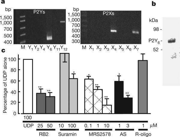
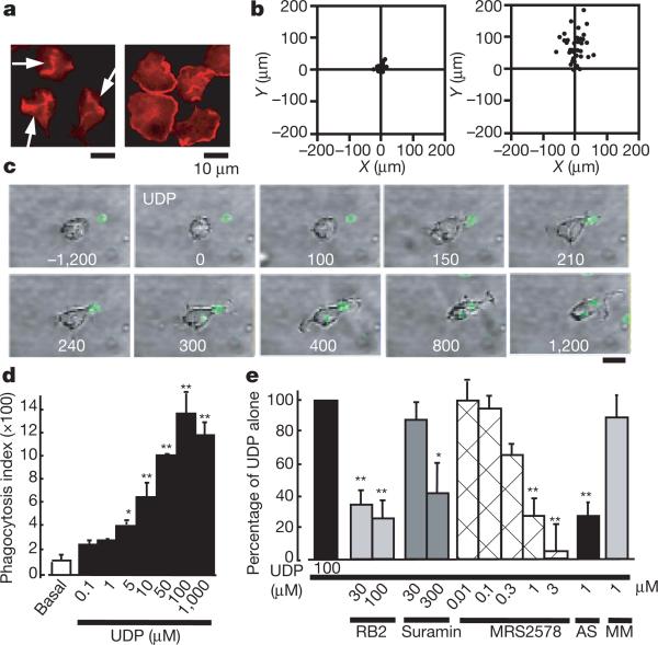
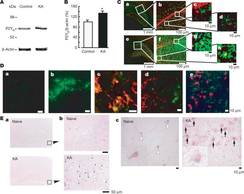
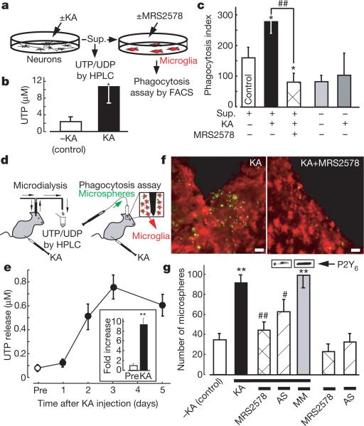
Microglia, brain immune cells, engage in the clearance of dead cells or dangerous debris, which is crucial to the maintenance of brain functions. When a neighbouring cell is injured, microglia move rapidly towards it or extend a process to engulf the injured cell. Because cells release or leak ATP when they are stimulated or injured, extracellular nucleotides are thought to be involved in these events. In fact, ATP triggers a dynamic change in the motility of microglia in vitro and in vivo, a previously unrecognized mechanism underlying microglial chemotaxis; in contrast, microglial phagocytosis has received only limited attention. Here we show that microglia express the metabotropic P2Y6 receptor whose activation by endogenous agonist UDP triggers microglial phagocytosis. UDP facilitated the uptake of microspheres in a P2Y6-receptor-dependent manner, which was mimicked by the leakage of endogenous UDP when hippocampal neurons were damaged by kainic acid in vivo and in vitro. In addition, systemic administration of kainic acid in rats resulted in neuronal cell death in the hippocampal CA1 and CA3 regions, where increases in messenger RNA encoding P2Y6 receptors that colocalized with activated microglia were observed. Thus, the P2Y6 receptor is upregulated when neurons are damaged, and could function as a sensor for phagocytosis by sensing diffusible UDP signals, which is a previously unknown pathophysiological function of P2 receptors in microglia.


Figures

Figure 1
Figure 1. Expression of P2Y6 receptor and UDP-evoked increase in [Ca2+]i in cultured microglia
a, RT–PCR analysis of the expression of mRNAs for P2Y6, P2Y12, P2X4 and P2X7 receptors in microglial cells. b, Expression of P2Y6 receptor protein confirmed by western blotting analysis. c, Effects of various chemicals on the increase in [Ca2+]i (measured as the change in ratio of fluorescence at 340 nm to that at 380 nm) evoked by 100 μM UDP in microglia. The maximum increase in Fura-2 fluorescence evoked by 100 μM UDP was considered as the control response, and values are expressed as a percentage of control. Data show means and s.e.m. for 24–36 cells obtained from at least three independent experiments. Significant differences from the response to UDP alone: asterisk, P < 0.05; two asterisks, P < 0.01 (Student's t-test).
Figure 2
Figure 2. Changes in cell motilities of microglia
a, UDP- and ATP-evoked membrane ruffles. Cultured microglia were stimulated for 5 min with 100 μM UDP (left) and 10 μM ATP (right), fixed, permeabilized, and then stained with anti-phalloidin. Scale bar, 10 μm. b, Typical chemotactic responses of microglia towards 100 μM UDP (left) and 100 μM ATP (right) assessed by the Dunn chemotaxis chamber (see Methods). c, Time-lapse images showing the effect of UDP on the microglial morphogenic changes and the uptake of fluorescent zymosan particles (green). The time after addition of UDP is shown in seconds in each picture. d, The UDP-evoked uptake of microspheres was assessed quantitatively as a phagocytosis index by using FACS. Data are mean and s.e.m. for three experiments (asterisk, P < 0.05; two asterisks, P < 0.01 compared with basal). e, Effects of the P2 receptor antagonists reactive blue 2 and suramin, the P2Y6 receptor antagonist MRS2578, and P2Y6 AS or MM on the UDP-evoked phagocytosis. Data are means and s.e.m. for three or four experiments (asterisk, P < 0.05; two asterisks, P < 0.01 compared with UDP alone).
Figure 3
Figure 3. Increase in P2Y6 receptors in the hippocampus after kainic acid (KA)-treatment
A, Western blot analysis, showing increase in P2Y6 receptor protein in rats treated intraperitoneally with 10 mg kg–1 KA, 72 h after treatment. B, Summary of quantitative data; KA was applied at 10 mg kg–1. Results are means and s.e.m. for 8 (control) and 7 (KA) experiments (asterisk, P < 0.05 compared with control). C, Immunohistochemical analysis in naive control (ad) and KA-treated (eh) rats; red, anti-NeuN antibody; green, anti-Iba1 antibody. Rectangles in a and e are expanded in b and f, respectively. Rectangles in b and f also correspond to c, d and g, h, respectively. D, Anti-P2Y6 antibody signals (green) were increased by KA (a, control; b, KA), which was colocalized with microglia (red in c, anti-OX42) but not with astrocytes (red in d, anti-GFAP) or neurons (red in e, anti-NeuN). E, ISH analysis. a, b, mRNA coding for P2Y6 receptor in naive rats was very low but was increased at the hippocampal CA3 region by KA (3 days later) (blue dots and arrowheads, KA). c, Double staining with P2Y6 antisense RNA probe (blue dots) and anti-Iba-1 antibody (brown signals, white (naive) or black (KA) arrows). In KA-treated rats there was an increased number of microglia, which was associated with P2Y6 receptor mRNA (blue signals, inset at higher magnification in KA).
Figure 4
Figure 4. KA-evoked increases in extracellular uridine nucleotides and P2Y6-receptor-mediated microglial phagocytosis in vitro and in vivo
a, Schematic diagram of the experiments in vitro. Sup., supernatant. b, Summary of the UTP concentration in the KA-treated and control supernatants. Data show means and s.e.m. for at least five independent experiments (asterisk, P < 0.05 compared with control). c, Effects of the KA-treated and control supernatant on microglial uptake of fluorescent latex beads. Data show means and s.e.m. for at least four independent experiments (asterisk, P < 0.05 compared with control; hash sign, P < 0.05 compared with KA-treated supernatant). d, Schematic diagram of the experiments in vivo. KA was applied intraperitoneally at 10 mg kg–1. e, Time course of changes in [UTP]o in baseline dialysates (before treatment with KA (Pre), and 1, 2, 3 and 5 days afterwards). Inset, fold increase at day 3 (compared with before treatment). f, Typical pictures of fluorescent microspheres (green) attached or taken up by microglia (red, anti-Iba1) in the KA-treated (left) and KA + MRS2578-treated (right) hippocampal CA3 regions. Scale bar, 20 μm. g, Quantitative analysis of phagocytosis in vivo (details are provided in Supplementary Methods). Changes in P2Y6 receptor protein by P2Y6 AS or MM are shown at the top of corresponding columns. Values are means and s.e.m. (asterisk, P < 0.01 compared with control (–KA); hash sign, P < 0.05; two hash signs, P < 0.01 compared with KA-treated group). Statistical analyses were performed by ANOVA with Scheffe's multiple comparison. At least three sections containing the injection sites were analysed per animal, and at least three animals were used in each group for analysis.

Comment in

References

    1. Guthrie PB, et al. ATP released from astrocytes mediates glial calcium waves. J. Neurosci. 1999;19:520–528. - PMC - PubMed
    1. Koizumi S, Fujishita K, Tsuda M, Shigemoto-Mogami Y, Inoue K. Dynamic inhibition of excitatory synaptic transmission by astrocyte-derived ATP in hippocampal cultures. Proc. Natl Acad. Sci. USA. 2003;100:11023–11028. - PMC - PubMed
    1. Nimmerjahn A, Kirchhoff F, Helmchen F. Resting microglial cells are highly dynamic surveillants of brain parenchyma in vivo. Science. 2005;308:1314–1318. - PubMed
    1. Davalos D, et al. ATP mediates rapid microglial response to local brain injury in vivo. Nature Neurosci. 2005;8:752–758. - PubMed
    1. Honda S, et al. Extracellular ATP or ADP induce chemotaxis of cultured microglia through Gi/o-coupled P2Y receptors. J. Neurosci. 2001;21:1975–1982. - PMC - PubMed

Publication types

MeSH terms