. 2007 May 2;26(9):2432-42.
doi: 10.1038/sj.emboj.7601672. Epub 2007 Apr 5.

The restriction fold turns to the dark side: a bacterial homing endonuclease with a PD-(D/E)-XK motif

Affiliations

The restriction fold turns to the dark side: a bacterial homing endonuclease with a PD-(D/E)-XK motif

Lei Zhao et al. EMBO J. .

Abstract

The homing endonuclease I-Ssp6803I causes the insertion of a group I intron into a bacterial tRNA gene-the only example of an invasive mobile intron within a bacterial genome. Using a computational fold prediction, mutagenic screen and crystal structure determination, we demonstrate that this protein is a tetrameric PD-(D/E)-XK endonuclease - a fold normally used to protect a bacterial genome from invading DNA through the action of restriction endonucleases. I-Ssp6803I uses its tetrameric assembly to promote recognition of a single long target site, whereas restriction endonuclease tetramers facilitate cooperative binding and cleavage of two short sites. The limited use of the PD-(D/E)-XK nucleases by mobile introns stands in contrast to their frequent use of LAGLIDADG and HNH endonucleases - which in turn, are rarely incorporated into restriction/modification systems.


Figures

Figure 1
Figure 1
Sequence alignment between the bacterial I-Ssp6803I homing endonuclease and archaeal Holliday junction resolvases. Only the first 110 residues of I-SspI, that align well, are shown with the homologous regions of the Hjc sequences. The final 40 residues of I-Ssp6803I that are not shown to participate in structural elaborations on the PD-(D/E)-XK core fold that are unique to I-SspI. Secondary structure elements of the homing endonuclease are shown above the alignment; structural elements from Pyrococcus furiosus Hjc are shown below. All of these elements are conserved in I-SspI with the exception of α-helix 2 (α2), which instead is an extended loop (‘L1' in the text and subsequent figures) that contacts the DNA target site. The blue stars above the alignment indicate conserved residues at active sites of the Hjc resolvase family. Residue labels in parentheses above the sequence alignment indicate mutations present in the crystal structure, as described in the text. Sequence alignments were carried out by ESPript (Gouet et al, 1999).
Figure 2
Figure 2
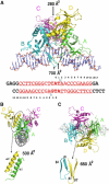
Structure of I-SspI bound to its DNA target site. Protein subunits are each colored separately. The complex is shown in three mutually orthogonal orientations in (AC). The buried surface area in each subunit interface is indicated. Two bound calcium ions are shown as red spheres. Loop L1 from monomers A and B is indicated by double black arrows. These loops are primarily associated with the central bases of the target site. These two loops do not interact with the bases in an identical manner—reflecting the asymmetry of this region of an otherwise symmetric target site. The same loops are disordered in subunits C and D, which are not bound to DNA and display minimal subunit contacts. The crystallization oligonucleotide construct is shown below panel A. The cleavage sites are indicated by cyan triangles. The base positions corresponding to physiological homing site are shown in red and the central 3-bases (corresponding both to the 3′ overhangs produced by cleavage and to anticodon triplet for fMet) are bold. The palindromic base pairs in the structure are underlined.
Figure 3
Figure 3
Structural comparison of protein subunits from the I-Ssp6803I homing endonuclease, the Hjc Holliday junction resolvase and the PvuII restriction endonuclease. (A) Structure and topology diagram of a single homing endonuclease subunit. The secondary structural elements are labeled and colored as follows: the PD-(D/E)-XK catalytic core region is pink and peripheral elaborations on that core are green. The N- and C-terminal residues of the secondary structural elements are indicated in the topology diagram. Catalytic residues are shown as sticks in the model on the left and labeled in red on the right. Regions involved in DNA recognition are indicated by dotted boxes and are numbered as shown in Figure 6 and described in the text. (B) The Hjc Holliday junction resolvase subunit. This structure has not been determined in the presence of DNA. (C) The PvuII restriction endonuclease subunit. Inlay: superposition of the I-SspI and Hjc catalytic cores (r.m.s.d. 1.9 Å).
Figure 4
Figure 4
Superposition of DNA-bound and unbound subunits in I-SspI. The endonuclease subunits are colored as in Figure 2. The DNA-bound subunits (A and B) and their bound DNA ligand are superimposed on subunit (C) of the two DNA-free monomers. As discussed in the text, this analysis indicates that the unbound subunits display a rigid-body rotational difference in their relative orientations and packing, as compared to the DNA-bound subunits. This results in a difference of approximately 6 Å in the position of the DNA-binding surface of subunit D relative to subunit A. The observed structure and packing of the C/D subunits cannot be accomodated for DNA binding either by DNA straightening (because of steric crowding in the central minor groove) or by repacking of subunit D (which would destabilize the A/D dimer interface).
Figure 5
Figure 5
Topology of I-Ssp6803I tetramer assembly and comparison with the SfiI restriction endonuclease. (A) Active sites and overall tetrameric packing of the I-Ssp6803I homing endonuclease. (B) Active sites and overall tetrameric packing of the SfiI restriction endonuclease. These two endonuclease have the same core fold and similar cleavage patterns (producing complementary 3-base, 3′ overhangs, as a result of cleavage across the minor groove) as shown below the models. Active sites from different monomers are colored in cyan or green (only secondary structures carrying the active site residues are shown for clarity). The cleavage sites on the DNA are indicated by red spots. The general architecture of the tetrameric assembly is indicted by the cartoon blocks representation, and are colored according to the corresponding protein subunits. The ribbon diagrams are shown with the ‘B' subunit from each structure in roughly similar orientations, to facilitate direct comparison of the tetrameric packing of the endonucleases.
Figure 6
Figure 6
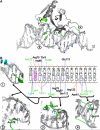
DNA-binding by I-SspI. (A) A single I-SspI monomer in complex with a DNA half-site. The regions in direct contact with bases are colored in green. Each distinct contact region on the protein is designated by numbers that correspond to Figure 3 and the text. These regions are magnified to show details in panel B. (B) Schematic diagram of DNA-binding and close-up views of the corresponding contacts. Only half of the DNA target is represented. Residues contacting DNA bases or backbone are labeled as follows: across the DNA, contacts in the minor groove are indicated on the left of each base while contacts in the major groove are indicated on their right. For the protein, residues observed making identical contacts in both monomers (A and B) are labeled in black, whereas residues observed making contacts in individual monomers A and B are labeled in green and blue, respectively. Contacts made by protein to DNA are colored as follows: blue lines indicate direct contacts between bases and protein side chains, blue dashed lines indicate direct contacts between bases and protein main chains, and red lines indicate nonspecific contacts to DNA backbone.
Figure 7
Figure 7
The active site of I-Ssp6803I. (A) The active site of I-Ssp6803I is shown as a ball-and-stick representation. The observed calcium ion position is shown as a red sphere. The anomalous difference map calculated from a native data set collected on a rotating anode X-ray source (CuKα; λ=1.54 Å) is shown in blue and contoured at 4.5σ. The predicted location of the water nucleophile and direction of its attack is indicated by the arrow; the scissile phosphodiester bond is indicated with a red star. (B) Superimposed active sites of I-SspI with EcoRV and Hjc. K51, D36 and E11 are conserved; Q49 is replaced by D and E, respectively in EcoRV and Hjc.

References

    1. Belfort M, Perlman PS (1995a) Mechanisms of intron mobility. J Biol Chem 270: 30237–30240 - PubMed
    1. Belfort M, Reaban ME, Coetzee T, Dalgaard JZ (1995b) Prokaryotic introns and inteins: a panoply of form and function. J Bacteriol 177: 3897–3903 - PMC - PubMed
    1. Belfort M, Roberts RJ (1997) Homing endonucleases: keeping the house in order. Nucleic Acids Res 25: 3379–3388 - PMC - PubMed
    1. Belle A, Landthaler M, Shub D (2002) Intronless homing: site-specific endonuclease SegF of bacteriophage T4 mediates localized marker exclusion analogous to homing endonucleases of group I introns. Genes Dev 16: 351–362 - PMC - PubMed
    1. Biniszkiewicz D, Cesnaviciene E, Shub DA (1994) Self-splicing group I intron in cyanobacterial initiator methionine tRNA: evidence for lateral transfer of introns in bacteria. EMBO J 13: 4629–4635 - PMC - PubMed

Associated data