Review
. 2007 Jul;36(7):609-26.
doi: 10.1007/s00256-006-0270-6. Epub 2007 Apr 5.

The spine in Paget's disease

Affiliations
Review

The spine in Paget's disease

C Dell'Atti et al. Skeletal Radiol. 2007 Jul.

Abstract

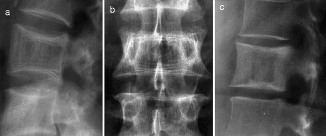
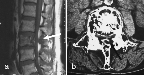
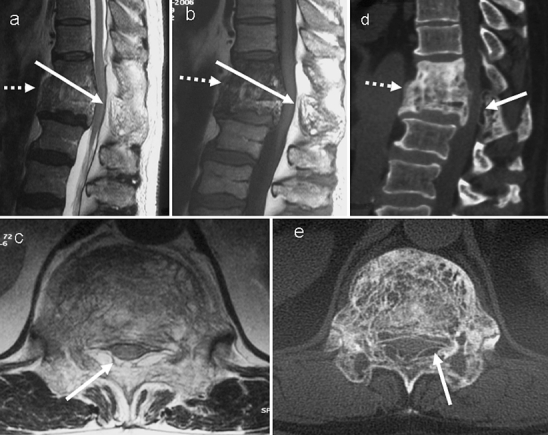
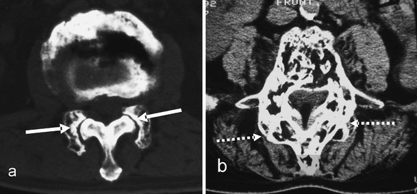
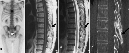
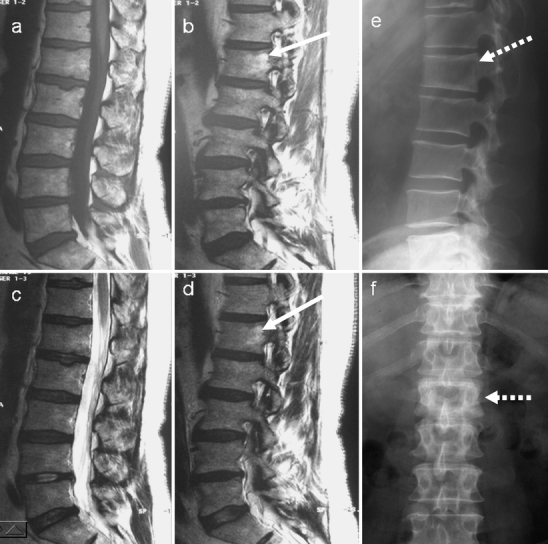
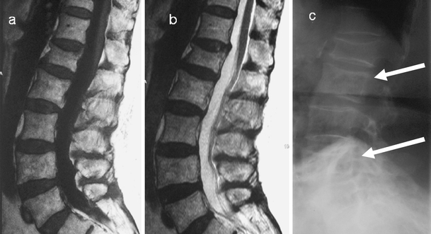
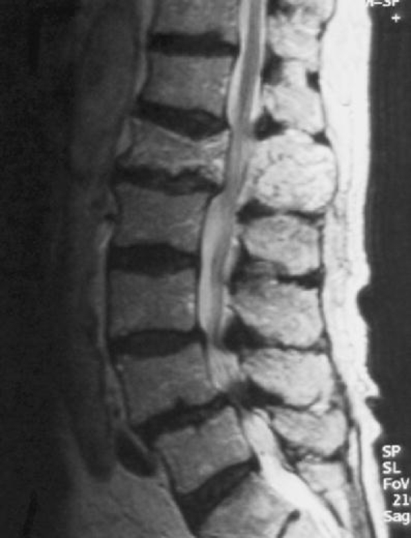
Paget's disease (PD) is a chronic metabolically active bone disease, characterized by a disturbance in bone modelling and remodelling due to an increase in osteoblastic and osteoclastic activity. The vertebra is the second most commonly affected site. This article reviews the various spinal pathomechanisms and osseous dynamics involved in producing the varied imaging appearances and their clinical relevance. Advanced imaging of osseous, articular and bone marrow manifestations of PD in all the vertebral components are presented. Pagetic changes often result in clinical symptoms including back pain, spinal stenosis and neural dysfunction. Various pathological complications due to PD involvement result in these clinical symptoms. Recognition of the imaging manifestations of spinal PD and the potential complications that cause the clinical symptoms enables accurate assessment of patients prior to appropriate management.


Figures

Fig. 1
Fig. 1
Diagram depicting the osseous mechanisms involved in vertebral body enlargement in Paget’s disease and its effect on the size of the marrow (dashed arrows) and cortex (solid arrows). A normal vertebra is depicted in the centre of the figure. a Periosteal apposition, normal endosteum resulting in thickened cortex, but with normal marrow size. b Periosteal apposition, endosteal resorption results in normal cortical thickness and an increased marrow size. c Periosteal apposition/endosteal apposition results in a thickened cortex and reduced marrow size. d Focal periosteal apposition results in a focal “pumice stone”-like enlargement
Fig. 2
Fig. 2
Axial CT sections in different patients showing the various mechanisms described in Fig. 1 and their effect on marrow size (long white arrow) and cortical thickness (short white arrow). a Periosteal apposition, normal endosteum. b Periosteal apposition, endosteal resorption. c Periosteal and endosteal apposition. d Pumice stone type (dashed arrow) of focal periosteal apposition. Similar focal periosteal apposition of the spinous process is seen
Fig. 3
Fig. 3
Diagram showing the periosteal and endosteal Pagetic osseous mechanisms involving the cortex of the spinal canal resulting in spinal canal narrowing. Normal cortical thickness (orange) of the spinal canal (white) is depicted at the top. a Expansion of bone due to periosteal apposition/endosteal resorption results in a thin cortical outline (solid black arrow) of the narrowed spinal canal (dashed arrow). b Bony expansion due to periosteal apposition/endosteal apposition results in a thickened cortical outline (solid black arrow) of the narrowed spinal canal (dashed arrow)
Fig. 4
Fig. 4
Axial CT images demonstrating the mechanisms in the posterior neural arch described in Fig. 3 and their effect on cortical thickness (solid arrow) and marrow size (dashed arrow). a Periosteal apposition/endosteal resorption. b Periosteal apposition/endosteal apposition
Fig. 5
Fig. 5
Vertebral Paget’s disease (PD) without expansion in two different patients. a PD of all the cervical vertebrae except C6. Note the absence of vertebral enlargement. There is sclerosis and loss of cortico-medullary differentiation of the vertebral bodies and the neural arches. b antero-posterior, c lateral radiographs and d axial CT through the L1 vertebra demonstrating sclerotic vertebra with no enlargement. The axial CT demonstrates trabecular and endosteal apposition, but no periosteal apposition accounting for the absence of enlargement. Diagnosis can be difficult and a biopsy (c) may be necessary in these cases
Fig. 6
Fig. 6
a Lateral and b antero-posterior radiographs demonstrate expansion of the vertebra with characteristic sclerotic lines parallel to the end-plates due to trabecular hypertrophy, an “early” sign of PD. c Lateral radiograph in a different patient demonstrates the “picture frame” vertebra due to thickening of the cortex and trabecular hypertrophy at the end-plates
Fig. 7
Fig. 7
a Sagittal T1-weighted MR image demonstrates PD in the L3 vertebral body and the posterior neural arch. The low T1 signal intensity mass (arrow) replacing epidural fat can be confused with epidural ossification. b However, an axial CT image confirms this to be due to expansion of the pagetic neural arch and not to ossification of the epidural fat. There is preservation of the intra-osseous fat as seen on both the MR and CT images, a useful discriminant from malignant infiltration
Fig. 8
Fig. 8
Paget’s disease of T11 and T12 showing an increased amount of high MR signal (solid white arrow) in the posterior epidural space at these levels on a sagittal T2-weighted, b sagittal T1-weighted and c axial T1-weighted images. This can be mistaken for epidural lipomatosis. However, the d CT sagittal and e axial images demonstrate this to be due to the fat density (solid white arrow) within the expanded posterior neural arch involved in PD. The axial images (c,e) were obtained at the level of the tip of the solid arrows on sagittal images. There is also fusion of the vertebrae (dashed arrow) across the intervertebral disc. The combination of anterior and posterior vertebral involvement in this case resulted in severe spinal canal narrowing and cord compression. Note the high T1 signal indicating a high fatty marrow content within the pagetic T11 and T12 vertebrae
Fig. 9
Fig. 9
Facet involvement. a Axial CT section demonstrates incongruity at the facet joint (solid white arrow) due to an involved enlarged facet of one vertebra articulating with an uninvolved non-expanded adjacent vertebral facet. b Axial CT section in a different patient demonstrates advanced facet joint arthropathy (dashed white arrows) across two pagetic facets with complete loss of joint space and new bone formation contributing to spinal canal stenosis. This can progress to fusion across the facet joints
Fig. 10
Fig. 10
a Initial scintigraphy for back pain demonstrates isolated increased uptake at a single vertebral level (T8). On initial inspection sagittal b T1-weighted and c T2-weighted MR images do not show any abnormality of the vertebral body. There is, though, some abnormal low signal from the posterior elements (black arrow). The diagnosis is still not clear. d However, a CT scan demonstrates the clear posterior vertebral (black arrow) sclerotic changes consistent with PD. Even on CT there are only minimal changes in the vertebral body
Fig. 11
Fig. 11
On initial examination, a sagittal and b parasagittal T1-weighted, c sagittal and d parasagittal T2-weighted MR images of the lumbar spine do not demonstrate any obvious abnormality. e, f The radiographs, however, show classic pagetic changes of the L1 vertebra (dashed arrows) including vertebral expansion, sclerosis and cortical thickening. Review of the MRI shows some minor increased signal in the expanded L1 vertebral body on both T1 and T2 parasagittal images, suggestive of fatty marrow change (white arrows)
Fig. 12
Fig. 12
a T1-weighted, b T2-weighted sagittal images of the lumbar spine demonstrate no marrow abnormality. There is only a subtle antero-posterior expansion of the L2 and L4. The diagnosis in these patients can be missed on initial MRI. c Lateral radiograph of the lumbar spine demonstrates the classic pagetic changes including vertebral expansion, trabecular hypertrophy and cortical thickening in L2 and L4. There is an incidental non-pagetic vertebral compression at L1. There is again preservation of the fat signal within the vertebrae involved in PD
Fig. 13
Fig. 13
Sagittal T2-weighted MR image demonstrates cauda equina compression at the L1 level due to pagetic enlargement of the whole vertebra. Note the stenosis caused by expansion of both the vertebral body and posterior elements. Degenerative spondylolisthesis and stenosis at L4/L5 is noted
Fig. 14
Fig. 14
Post-myelographic lateral lumbar spine a radiograph and b axial CT scan demonstrate spinal block at the L1 vertebral level, which is pagetic. Note the severe degree of spinal stenosis despite the apparent increase in the interpedicular distance on the axial CT image
Fig. 15
Fig. 15
Sagittal fat suppressed T1-weighted image after gadolinium-DTPA administration in the same patient as in Fig. 8 demonstrates intense enhancement at both the T12/L1 and the L3/L4 levels (arrows). The enhancement in part reflects increased blood supply to the vertebra
Fig. 16
Fig. 16
Lateral and anteroposterior radiograph of the lumbar spine demonstrates severe compression of a pagetic L3 vertebra with retropulsion into the spinal canal. Note the increased interpediculate distance as a hallmark of PD
Fig. 17
Fig. 17
Lateral radiographs in two different patients with PD showing two different mechanisms of spondylolisthesis: a spondylolytic (arrow) spondylolisthesis; b degenerative spondylolisthesis
Fig. 18
Fig. 18
Disc involvement. Serial radiographs 2 years apart in the same patient demonstrate progressive involvement of the L4/L5 intervertebral disc in Paget’s disease of the L5
Fig. 19
Fig. 19
Extra-osseous Paget’s disease. a Sagittal CT reconstruction and b T1-weighted MR sagittal image demonstrate anterior longitudinal ligament ossification (white arrows). c, d Axial CT sections demonstrate ossification in the ligamentum flavum due to extra-osseous PD (black arrows) in a different patient. e Antero-posterior radiograph in another patient demonstrates paravertebral soft tissue swelling (dashed arrows) producing the “pseudo-sarcoma” appearance
Fig. 20
Fig. 20
Vertebral metastasis from colon carcinoma. Sagittal a T1- and b T2-weighted MR images demonstrate metastasis in L2 and L4 vertebral bodies seen as discrete lesions (white arrows) with low signal on T1- and high signal on T2-weighted images. A further epidural lesion (black arrow) is seen in the spinal canal posteriorly at L3. Note the pagetic changes with expansion of L2 and L3 vertebral bodies

References

    1. {'text': '', 'ref_index': 1, 'ids': [{'type': 'DOI', 'value': '10.1016/S0140-6736(56)90422-6', 'is_inner': False, 'url': 'https://doi.org/10.1016/s0140-6736(56)90422-6'}, {'type': 'PubMed', 'value': '13347083', 'is_inner': True, 'url': 'https://pubmed.ncbi.nlm.nih.gov/13347083/'}]}
    2. Collins DH. Paget’s disease of bone; incidence and subclinical forms. Lancet 1956; 271 6933: 51–57. - PubMed
    1. {'text': '', 'ref_index': 1, 'ids': [{'type': 'DOI', 'value': '10.1007/BF01887990', 'is_inner': False, 'url': 'https://doi.org/10.1007/bf01887990'}]}
    2. Schmorl G. Über Osteitis deformans Paget. Virchows Arch Pathol Anat Physiol 1932; 238: 694–751.
    1. {'text': '', 'ref_index': 1, 'ids': [{'type': 'PubMed', 'value': '3103963', 'is_inner': True, 'url': 'https://pubmed.ncbi.nlm.nih.gov/3103963/'}]}
    2. Maldague B, Malghem J. Dynamic radiologic patterns of Paget’s disease of bone. Clin Orthop Relat Res 1987; 217: 126–151. - PubMed
    1. {'text': '', 'ref_index': 1, 'ids': [{'type': 'PubMed', 'value': '3549098', 'is_inner': True, 'url': 'https://pubmed.ncbi.nlm.nih.gov/3549098/'}]}
    2. Basle MF, Rebel A, Fournier JG, Russell WC, Malkani K. On the trail of paramyxoviruses in Paget’s disease of bone. Clin Orthop Relat Res 1987; 217: 9–15. - PubMed
    1. {'text': '', 'ref_index': 1, 'ids': [{'type': 'DOI', 'value': '10.1007/s005860100329', 'is_inner': False, 'url': 'https://doi.org/10.1007/s005860100329'}, {'type': 'PMC', 'value': 'PMC3611523', 'is_inner': False, 'url': 'https://pmc.ncbi.nlm.nih.gov/articles/PMC3611523/'}, {'type': 'PubMed', 'value': '11718191', 'is_inner': True, 'url': 'https://pubmed.ncbi.nlm.nih.gov/11718191/'}]}
    2. Hadjipavlou AG, Gaitanis LN, Katonis PG, Lander P. Paget’s disease of the spine and its management. Eur Spine J 2001; 10 5: 370–384. - PMC - PubMed

MeSH terms