. 2007 Apr;3(4):e49.
doi: 10.1371/journal.ppat.0030049.

M. tuberculosis Ser/Thr protein kinase D phosphorylates an anti-anti-sigma factor homolog

Affiliations

M. tuberculosis Ser/Thr protein kinase D phosphorylates an anti-anti-sigma factor homolog

Andrew E Greenstein et al. PLoS Pathog. 2007 Apr.

Abstract

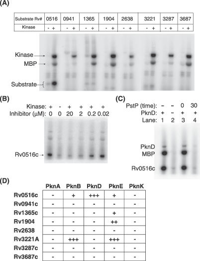
Receptor Ser/Thr protein kinases are candidates for sensors that govern developmental changes and disease processes of Mycobacterium tuberculosis (Mtb), but the functions of these kinases are not established. Here, we show that Mtb protein kinase (Pkn) D overexpression alters transcription of numerous bacterial genes, including Rv0516c, a putative anti-anti-sigma factor, and genes regulated by sigma factor F. The PknD kinase domain directly phosphorylated Rv0516c, but no other sigma factor regulator, in vitro. In contrast, the purified PknB and PknE kinase domains phosphorylated distinct sigma regulators. Rather than modifying a consensus site, PknD phosphorylated Rv0516c in vitro and in vivo on Thr2 in a unique N-terminal extension. This phosphorylation inhibited Rv0516c binding in vitro to a homologous anti-anti-sigma factor, Rv2638. These results support a model in which signals transmitted through PknD alter the transcriptional program of Mtb by stimulating phosphorylation of a sigma factor regulator at an unprecedented control site.


Conflict of interest statement

Competing interests. The authors have declared that no competing interests exist.

Figures

Figure 1
Figure 1. Efficient Overexpression of PknD and Concomitant Induction of Rv0516c in Logarithmically Growing Mtb
(A) Western blots showing kinase levels (anti-PknD) and kinase activity (anti-phosphothreonine) at 0, 2, 4, 8 and 24 h after induction of WT and Asp138Asn PknD (D138N). As a loading control, the amount of the KatG protein present at a constant level was monitored using anti-KatG antibodies. Addition of the inducer (acetamide) to WT Mtb (lacking an inducible PknD gene) caused no discernable change in the pattern of phosphoproteins (unpublished data). The presence of a band at the molecular weight of phosphorylated WT PknD at time zero may indicate that the kinase was already expressed (and autophosphorylated) at low levels in the absence of inducer. In contrast, phosphorylated Asn138Asn PknD accumulated only after the inducer was added. (B) Cluster diagram showing the eight genes with the largest time-dependent changes in transcript levels upon PknD induction. Expression levels are shown relative to a reference pool in which equal amounts of RNA from each sample were mixed and the value at time zero was normalized to 1.0. Microarray analysis revealed >2-fold induction or repression of 137 genes (Table S1). Among the eight most altered transcripts shown here, we observed robust induction (blue) of the putative anti-anti–sigma factor Rv0516c. Rv0516c expression increased much more upon expression of WT PknD than the attenuated mutant (Asp138Asn). Genes labeled in red are among those most reduced in expression during logarithmic phase growth of an Mtb sigF knockout mutant [21]. Color unit is fold change of gene expression.
Figure 2
Figure 2. Mtb Proteins Containing Domains Homologous to the B. subtilis Alternative Sigma Factor Regulators SpoIIAA and SpoIIAB
Four of the Mtb homologs of the B. subtilis SpoIIAB (anti–sigma factor) or SpoIIAA (anti-anti–sigma factor) encode proteins that have been shown to regulate SigF or SigH. Rv0941c contains both types of domain, and Rv1364c contains both domains as well as an RsbU-like, Ser/Thr phosphatase domain. Three of the seven anti-anti–sigma factor domains are preceded by a Ser/Thrrich N-terminal extension (blue). This extension in Rv0516c contains the site of PknD phosphorylation.
Figure 3
Figure 3. PknD Specifically Phosphorylates Rv0516c In Vitro
(A) Each anti–sigma factor or anti-anti–sigma factor (gene [Rv] number listed along the top) fused to MBP was cloned, expressed, purified, and incubated with (+) or without (−) PknD in a [γ-32P]ATP transfer assay. The assay was quenched with EDTA, and TEV protease was added to separate the MBP tag from the sigma factor regulator. The two dark bands in the center of each PknD lane correspond to the autophosphorylated kinase and the MBP tag. The sigma factor regulators released by TEV protease are between 12 and 20 kDa. Only Rv0516c was phosphorylated by PknD. (B) Dose-dependent attenuation by the PknD inhibitor, SP600125, shows that PknD phosphorylates Rv0516c. (C) PstP, a protein Ser/Thr phosphatase from Mtb, dephosphorylates Rv0516c. PknD (lane 1), but not heat-inactivated PknD (lane 2), phosphorylated Rv0516c. PstP was added to PknD-phosphorylated Rv0516c, and the phosphatase reaction was quenched immediately (lane 3) or after 30 min (lane 4). (D) In vitro kinase assays were performed to investigate the specificity of five Mtb kinases for the eight sigma factor regulators (Figure S3). Assays were carried out in a manner identical to the assay shown in Figure 3A. Kinase concentrations were adjusted to catalyze equal phosphorylation of myelin basic protein in vitro. PknB, PknD, and PknE phosphorylated a specific subset of substrates.
Figure 4
Figure 4. PknD Phosphorylates Rv0516c on Thr2 In Vitro
(A) Mass spectrometry of trypsin-digested, PknD-phosphorylated Rv0516c indicated the presence of a single phosphorylation site within the N-terminal nine residues. The figure shows the mass spectrum of an HPLC fraction of the digest. The measured mass (monoisotopic MH+) of the N-terminal peptide (1384.60 Da [arrow]) was 79.97 Da larger than the expected mass of 1,304.63 Da, corresponding to addition of a single phosphate group. Tandem MS and N-terminal sequencing of the peptide at m/z 1,384.60 revealed Thr2 as the site of phosphorylation. (B) Rv0516c Thr2 occurs in a unique, 28-residue Ser/Thr-rich, N-terminal extension, not in the anti-anti–sigma factor domain. (C) Mutations in the N-terminus of Rv0516c confirm that PknD phosphorylates Rv0516c on Thr2. The eight indicated mutants of Rv0516c were purified, and in vitro phosphorylation by the PknD kinase domain was assayed after cleavage with the TEV protease (left). Non-native residues before the start of Rv0516c left after TEV protease cleavage are shaded in gray. Protein-stained loading controls (center) confirmed that an equal amount of each Rv0516c variant was assayed. Controls lacking kinase showed that the phosphorylation was due to PknD (far right). Thr2Ala (T2A) and deletions of the N-terminal four and eight residues caused the most dramatic reductions in phosphorylation.
Figure 5
Figure 5. PknD Phosphorylates Rv0516c on Thr2 In Vivo, and This Phosphorylation Blocks Binding to Another Sigma Factor Regulator
(A) Western blot of lysates of Mtb strains expressing full-length PknD under the control of an acetamide-inducible promoter and constitutively expressing Rv0516c with a C-terminal, FLAG epitope tag. As controls, the kinase was rendered inactive (D138N) or the Rv0516c phosphorylation site was mutated (Thr2Ala [T2A]). The active (but not the mutant) PknD phosphorylated the overexpressed WT Rv0516c. The levels of endogenous proteins were too low to detect phosphorylated Rv0516c in this experiment. KatG was detected with antibodies as a loading control. Rv0516c (but not the Thr2Ala mutant) was phosphorylated upon WT PknD induction. (B) Phosphorylation on Thr2 abolished binding to Rv2638. Equal amounts of cell lysates overexpressing Rv2638 were incubated with pre-phosphorylated (lane 4) or unphosphorylated (lane 3) Rv0516c. As a control, Rv2638 binding to the purification tag alone was evaluated (lane 2). Protein staining was used to insure that equal amounts of recombinant protein were used as bait in each reaction, and bound Rv2638 was visualized by Western blotting with antibodies against the His6 tag. Total soluble protein before incubation with the amylose-Sepharose (lane 1) indicated the presence of Rv2638. Only unphosphorylated Rv0516c bound the anti-anti–sigma factor, Rv2638.

References

    1. Dye C, Scheele S, Dolin P, Pathania V, Raviglione MC. Consensus statement. Global burden of tuberculosis: Estimated incidence, prevalence, and mortality by country. WHO Global Surveillance and Monitoring Project. JAMA. 1999;282:677–686. - PubMed
    1. Zhang Y. Persistent and dormant tubercle bacilli and latent tuberculosis. Front Biosci. 2004;9:1136–1156. - PubMed
    1. Russell DG. Mycobacterium tuberculosis: Here today, and here tomorrow. Nat Rev Mol Cell Biol. 2001;2:569–577. - PubMed
    1. Cole ST, Brosch R, Parkhill J, Garnier T, Churcher C, et al. Deciphering the biology of Mycobacterium tuberculosis from the complete genome sequence. Nature. 1998;393:537–544. - PubMed
    1. Av-Gay Y, Everett M. The eukaryotic-like Ser/Thr protein kinases of Mycobacterium tuberculosis . Trends Microbiol. 2000;8:238–244. - PubMed

Publication types

MeSH terms