. 2007 Apr 6;316(5821):120-3.
doi: 10.1126/science.1136985.

An ATP gate controls tubulin binding by the tethered head of kinesin-1

Affiliations

An ATP gate controls tubulin binding by the tethered head of kinesin-1

Maria C Alonso et al. Science. .

Abstract

Kinesin-1 is a two-headed molecular motor that walks along microtubules, with each step gated by adenosine triphosphate (ATP) binding. Existing models for the gating mechanism propose a role for the microtubule lattice. We show that unpolymerized tubulin binds to kinesin-1, causing tubulin-activated release of adenosine diphosphate (ADP). With no added nucleotide, each kinesin-1 dimer binds one tubulin heterodimer. In adenylyl-imidodiphosphate (AMP-PNP), a nonhydrolyzable ATP analog, each kinesin-1 dimer binds two tubulin heterodimers. The data reveal an ATP gate that operates independently of the microtubule lattice, by ATP-dependent release of a steric or allosteric block on the tubulin binding site of the tethered kinesin-ADP head.


Figures

Fig. 1
Fig. 1
Activation of kinesin dimers by tubulin and microtubules. The microtubule (MT) or tubulin heterodimer (HD) stimulated steady state ATPase activity of kinesin was measured at 25°C using an enzyme-linked assay in 20 mM PIPES, pH 6.9, 5 mM MgCl2, 1mM DTT (Supplementary Material). Values for Vmax and Km were obtained by least squares fitting to plots of ATPase versus tubulin heterodimer concentration, using Kaleidagraph 3.6.4 (Synergy Software). (A) Vmax 38.1 s−1, Km 0.44 μM for heterodimers (HD), Vmax 36.4 s−1, Km 0.39 μM for microtubules (MT). (B) Vmax 0.9 s−1, Km 2.03 μM for HD, Vmax 12.2 s−1, Km 0.28 μM for MT. (C) Vmax 35.9 s−1, Km 1.29 μM for HD, Vmax 71.1 s−1, Km 0.62 μM for MT. (D) Vmax 4.0 s−1, Km 0.86 μM for HD, Vmax 17.0 s−1, Km 0.49 μM for MT.
Fig. 2
Fig. 2
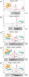
Superose 12 column chromatography of kinesin-tubulin complexes. (A) 6.5 μM rK430 rat kinesin. (B) 13 μM pig brain tubulin. (C) 13 μM tubulin + 6.5 μM rat kinesin; no added nucleotide. (D) 13 μM tubulin + 6.5 μM kinesin in 0.2 mM AMPPNP. (D) 13 μM tubulin + 6.5 μM kinesin in 2 mM ADP. Y axis marks are in mAU at 290 nm. X axis marks are at intervals of 1 ml. The included volume of the column was 20.0 ml and the void volume was 8.1 ml. 240μl samples were run at 0.5 ml min−1 in 50 mM PIPES pH 6.9, 2 mM MgCl2, 1 mM EGTA with or without 2 mM ADP or 0.2 mM AMPPNP. The grey vertical line indicates the tubulin elution position. The elution profiles of tubulin alone and kinesin alone were the same in ADP or AMPPNP. Binding stoichiometry was measured for the shaded fractions.
Fig. 3
Fig. 3
2-step tubulin-activated ADP release from kinesin. (A,C) Fluorescence transients corresponding to slow binding of 1 μM mantATP to 1 μM rat kinesin, followed by slow release of mantADP from both kinesin heads induced by a chase of nonfluorescent 1 mM ATP or 1 mM AMPPNP. (B,D) The same experiment but with 2 μM tubulin heterodimers added before the addition of the chasing nucleotide. Buffer 20 mM PIPES, pH 6.9, 2 mM MgCl2.
Fig. 4
Fig. 4
Fitting of cryoEM maps of the apo state of kinesin dimers attached to microtubules. The microtubule plus end is towards the top of the page. (Left) CryoEM map (20) (Right) Fitted orientation of two heads of rat kinesin. One head (yellow) is attached to the underlying microtubule protofilament, whilst the other head (orange) is parked in a forwards-biased position that masks its tubulin binding site.
Fig.5
Fig.5
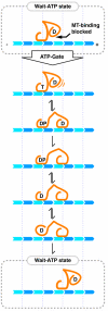
Gating scheme. The cycle begins with ATP-gated exit of the tethered head from a refractory dwell state (dotted box) in which the tubulin binding site of the tethered head is blocked. Following ATP binding, the block is released and the tethered head is then free to diffuse to its next site along the microtubule. Hydrolysis and phosphate release on the trailing head return it to a weak binding K.ADP state, which then detaches, diffuses to a forwards-biased position and reverts to a blocked state.

Comment in

References

    1. Howard J, Hudspeth AJ, Vale RD. Nature. 1989;342:154. - PubMed
    1. Hua W, Young EC, Fleming ML, Gelles J. Nature. 1997;388:390. - PubMed
    1. Kaseda K, Higuchi H, Hirose K. Nat Cell Biol. 2003;5:1079. - PubMed
    1. Asbury CL, Fehr AN, Block SM. Science. 2003;302:2130. - PMC - PubMed
    1. Yildiz A, Tomishige M, Vale RD, Selvin PR. Science. 2004;303:676. - PubMed

Publication types