. 2007 May;117(5):1314-23.
doi: 10.1172/JCI31222. Epub 2007 Apr 5.

Ewing sarcoma gene EWS is essential for meiosis and B lymphocyte development

Affiliations

Ewing sarcoma gene EWS is essential for meiosis and B lymphocyte development

Hongjie Li et al. J Clin Invest. 2007 May.

Abstract

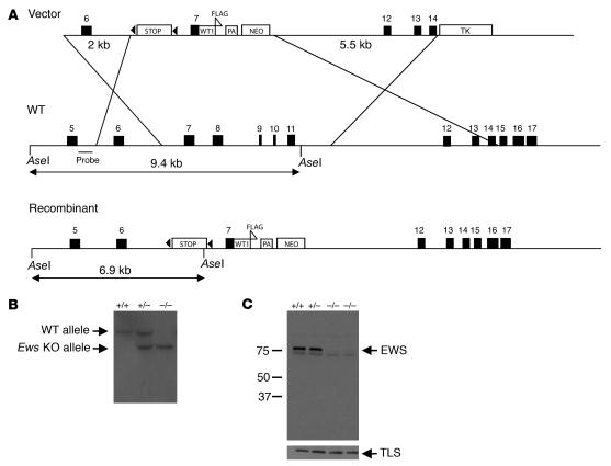
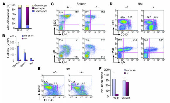
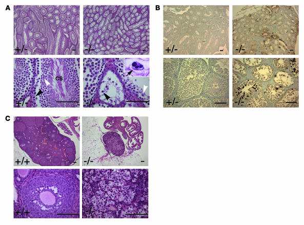
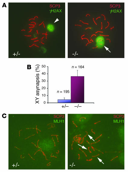
Ewing sarcoma gene EWS encodes a putative RNA-binding protein with proposed roles in transcription and splicing, but its physiological role in vivo remains undefined. Here, we have generated Ews-deficient mice and demonstrated that EWS is required for the completion of B cell development and meiosis. Analysis of Ews(-/-) lymphocytes revealed a cell-autonomous defect in precursor B lymphocyte (pre-B lymphocyte) development. During meiosis, Ews-null spermatocytes were deficient in XY bivalent formation and showed reduced meiotic recombination, resulting in massive apoptosis and complete arrest in gamete maturation. Inactivation of Ews in mouse embryonic fibroblasts resulted in premature cellular senescence, and the mutant animals showed hypersensitivity to ionizing radiation. Finally, we showed that EWS interacts with lamin A/C and that loss of EWS results in a reduced lamin A/C expression. Our findings reveal essential functions for EWS in pre-B cell development and meiosis, with proposed roles in DNA pairing and recombination/repair mechanisms. Furthermore, we demonstrate a novel role of EWS in cellular senescence, possibly through its interaction and modulation of lamin A/C.


Figures

Figure 1
Figure 1. Inactivation of Ews by gene targeting.
(A) Scheme for Ews targeting strategy. The triangles indicate loxP sequences flanking the stop cassette. WT1, WT1 cDNA; PA, poly(A) sequence. The probe used for Southern blot analysis is indicated. (B) Southern blot analysis of AseI-digested tail DNA. Wild-type (9.4-kb) and targeted (6.9-kb) alleles are marked. (C) Western blot analysis of EWS and TLS using whole-cell extracts isolated from MEFs. Ews genotype and the molecular weight markers (kDa) are indicated.
Figure 2
Figure 2. EWS is intrinsically required for pre–B cell development.
(A) A complete blood count was performed with peripheral blood from 3-week-old littermates (n = 3/genotype). White blood cell differential data indicate a marked reduction of the lymphocyte population in Ews–/– mice. (B) Reduced cellularity of lymphoid organs in Ews–/– mice. Total cell count is shown for indicated organs of 3-week-old mice (n = 3/genotype). Flow cytometry of splenocytes (C) and bone marrow–derived cells (D) from Ews–/– mice and littermate controls using B cell markers B220, CD43, IgM, and IgD. (E) Fetal liver chimera analysis. Bone marrow cells were harvested from fetal liver chimeras and analyzed by flow cytometry with antibodies against CD43 and B220 along with CD45.1 and CD45.2. Presented are representative data from 3 independent experiments with similar results (n = 3 for Ews–/– and n = 3 for Ews+/– littermate controls). (F) CFU assay. Bone marrow cells from fetal liver chimeras were plated in duplicate in either pre-B or multilineage differentiation (GM-CFU) media. The number of colonies is presented as the mean colony number from 3 independent experiments.
Figure 3
Figure 3. Ews–/– mice are defective in spermatogenesis and oogenesis.
(A) H&E staining of testes from 3-month-old Ews–/– and Ews+/– littermates. Black arrowheads mark primary spermatocytes, and white arrowheads indicate round spermatids. CS, condensing spermatids. The arrow in the inset shows a multinucleated spermatocyte. (B) TUNEL staining of testes from 3-month-old Ews–/– and Ews+/– littermates shows massive apoptosis of spermatocytes in the mutant testes. (C) H&E staining of ovaries from 3-month-old Ews–/– and Ews+/– littermates. Note the complete absence of oocytes and developing follicles in Ews–/– ovary (right panels). A minimum of 3 animals for each genotype were examined for H&E and TUNEL analyses. Scale bars: 100 μm.
Figure 4
Figure 4. Reduced meiotic recombination and XY asynapsis in Ews–/– spermatocytes.
(A) Double immunofluorescence staining of spermatocyte spread from 6-week-old testes with α-SCP3 and α-γH2AX antibodies. The arrowhead (left panel) shows a sex body containing bivalent XY, and the arrow (right panel) indicates univalent XY. Original magnification, ×40. (B) Univalent XY were counted under a fluorescence microscope and represented as the percentage of total pachytene nuclei examined (3 mice/genotype). n values represent total pachytene nuclei. (C) Double immunofluorescence staining of spermatocyte spread with α-SCP3 and α-MLH1 antibodies. The arrows in the right panel indicate bivalents with no MLH1 staining. Original magnification, ×40.
Figure 5
Figure 5. Premature cellular senescence and aging-like features in Ews–/– mice.
(A) MEFs were continuously passaged, and cell count determined at every passage is plotted. n = 3/genotype. (B) Senescence-associated β-galactosidase staining of passage 3 Ews+/+ and Ews–/– MEFs. A representative field is shown. Original magnification, ×20. (C) Cells positively stained for senescence-associated β-galactosidase activity were counted under a microscope. Results represent an average of 4 independent cell lines examined at passage 3 for each genotype. (D) Western blot analysis of p19, p21, p53, p16, pRb, EWS, and actin using 3 independently derived MEFs of each genotype. (E) Representative x-ray images of the whole body and the dissected long bones of littermates are shown. X-rays were taken of 3- to 6-week-old mice of the indicated genotypes (n = 4). (F) H&E staining of skin sections from a similar dorsal area of 3-week-old littermates. F, subcutaneous fat. M, muscle. Scale bars: 100 μm. (G) Kaplan-Meier survival curve of mice that received 7 Gy of ionizing radiation on day 1.
Figure 6
Figure 6. Endogenous interaction of EWS and lamin A/C.
(A) Localization of GFP-EWS and endogenous lamin A/C in PC3 cells by immunofluorescence microscopy. Arrow in the merged panel indicates the colocalization (yellow) in the inner nuclear membrane. Original magnification, ×40. (B) HeLa nuclear extracts immunoprecipitated with either preimmune (control) or EWS antibodies were immunoblotted with α-lamin A/C antibodies. WB, Western blot. (C) Immunofluorescence microscopy of endogenous lamin A/C in Ews+/– or Ews–/– MEFs. Original magnification, ×20. (D) Western blot analysis of MEFs with indicated genotypes using α-lamin A/C or α-actin antibodies. (E) Quantification of lamin A/C relative to actin in the immunoblot (D) using the Odyssey infrared system.

References

    1. Delattre O., et al. Gene fusion with an ETS DNA-binding domain caused by chromosome translocation in human tumours. Nature. 1992;359:162–165. - PubMed
    1. Arvand A., Denny C.T. Biology of EWS/ETS fusions in Ewing’s family tumors. Oncogene. 2001;20:5747–5754. - PubMed
    1. Janknecht R. EWS-ETS oncoproteins: the linchpins of Ewing tumors. Gene. 2005;363:1–14. - PubMed
    1. Zucman J., et al. EWS and ATF-1 gene fusion induced by t(12;22) translocation in malignant melanoma of soft parts. Nat. Genet. 1993;4:341–345. - PubMed
    1. Panagopoulos I., et al. Fusion of the EWS and CHOP genes in myxoid liposarcoma. Oncogene. 1996;12:489–494. - PubMed

Publication types

MeSH terms

Substances