. 2007 May;117(5):1324-34.
doi: 10.1172/JCI31240. Epub 2007 Apr 5.

Modulation of adverse cardiac remodeling by STARS, a mediator of MEF2 signaling and SRF activity

Affiliations

Modulation of adverse cardiac remodeling by STARS, a mediator of MEF2 signaling and SRF activity

Koichiro Kuwahara et al. J Clin Invest. 2007 May.

Abstract

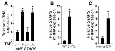
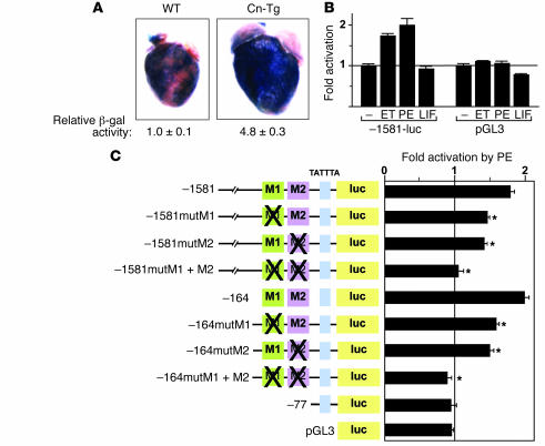
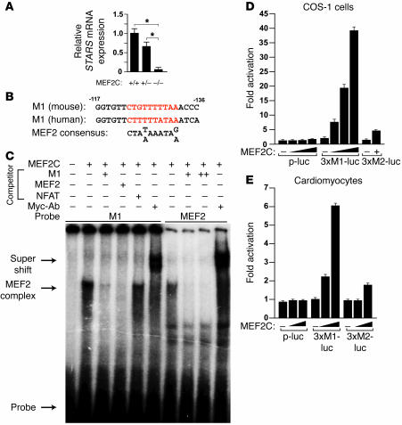
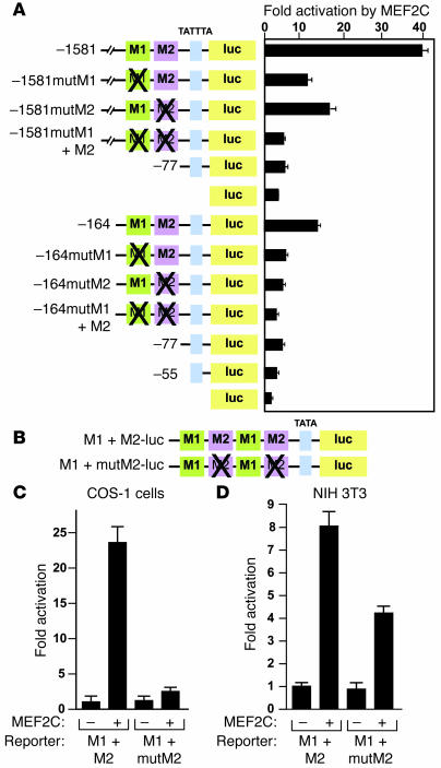
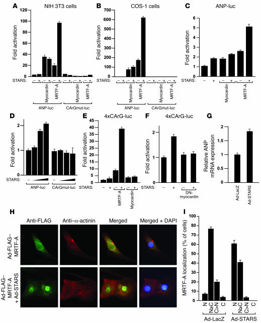
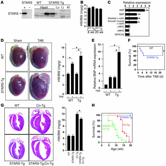
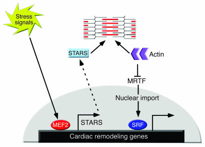
Cytoskeletal proteins have been implicated in the pathogenesis of cardiomyopathy, but how the cytoskeleton influences the transcriptional alterations associated with adverse cardiac remodeling remains unclear. Striated muscle activator of Rho signaling (STARS) is a muscle-specific actin-binding protein localized to the Z disc that activates serum response factor-dependent (SRF-dependent) transcription by inducing nuclear translocation of the myocardin-related SRF coactivators MRTF-A and -B. We show that STARS expression is upregulated in mouse models of cardiac hypertrophy and in failing human hearts. A conserved region of the STARS promoter containing an essential binding site for myocyte enhancer factor-2 (MEF2), a stress-responsive transcriptional activator, mediates cardiac expression of STARS, which in turn activates SRF target genes. Forced overexpression of STARS in the heart sensitizes the heart to pressure overload and calcineurin signaling, resulting in exaggerated deterioration in cardiac function in response to these hypertrophic stimuli. These findings suggest that STARS modulates the responsiveness of the heart to stress signaling by functioning as a cytoskeletal intermediary between MEF2 and SRF.


Figures

Figure 1
Figure 1. Upregulation of STARS during cardiac hypertrophy and failure as analyzed by real-time RT-PCR.
(A) Transcripts for STARS and ANP were detected in hearts of mice subjected to TAB and control sham operation. n = 6. (B) Expression of STARS mRNA in Cn-Tg mice and control non-Tg littermates (WT). n = 6. (C) Expression of STARS mRNA in human hearts with idiopathic cardiomyopathy (ICM) and control normal hearts. n = 3. *P < 0.05.
Figure 2
Figure 2. Mutational analysis of the STARS promoter.
(A) Fragments of the STARS upstream region linked to the hsp68 minimal promoter and lacZ were used to generate Tg mice. (B) LacZ staining of Tg mice at E12.5 carrying the indicated STARS-lacZ transgenes. H&E-stained sections of the heart are shown. (C) LacZ staining of striated muscle of Tg mice carrying STARS-lacZ construct 1. TA, tibialis anterior; EDL, extensor digitorum longis; sol, soleus. (D and E) Luciferase activity was measured in cardiomyocytes (D and E) and COS-1 cells (E) transfected with the luciferase reporter plasmid pGL3 linked to upstream fragments of the STARS gene. (F) Sequences of the M1 and M2 regions with mutations shown in red. (G) Luciferase activity was measured in cardiomyocytes transfected with –1581- or –164STARS-luc without or with mutations in the M1 and/or M2 region (mutM1, mutM2, mutM1+M2). *P < 0.05 versus reporter without mutation. (H) LacZ staining of E12.5 Tg embryos carrying wild type construct 1 or construct 1 with mutations in the M1 and M2 regions (mutM1+M2). Original magnification, ×2 (B, upper panels, and H); ×10 (B, lower panels).
Figure 3
Figure 3. The proximal promoter region mediates inducible expression of the STARS gene.
(A) LacZ staining and activity (n = 4) of adult Tg mouse hearts carrying construct 1 in wild-type or Cn-Tg mice. Original magnification, ×2. (B) Luciferase activity of cardiomyocytes transfected with –1581STARS-Luc or control pGL3 treated with 100 nM ET, 100 μM PE, or 1 nM leukemia inhibitory factor (LIF) for 24 hours. (C) Luciferase activity of cardiomyocytes transfected with –1581- or –164STARS-luc with or without mutations in the M1 or M2 regions and treated with 100 μM PE for 24 hours. *P < 0.05 versus the reporter without mutation.
Figure 4
Figure 4. Regulation of the STARS promoter by MEF2.
(A) STARS mRNA expression in wild-type, Mef2c+/–, and Mef2c–/– embryos at E8.5 (n = 3). (B) Sequences of mouse and human M1 region aligned with MEF2 consensus binding site. Sequences are on the opposite strand relative to those in Figure 2F. (C) Gel mobility shift assay using in vitro translated myc-tagged MEF2C with radiolabeled STARS M1 DNA sequence or desmin MEF2-binding site as probe. Nonlabeled M1, desmin MEF2, or an unrelated NFAT consensus site was used as competitor (+ denotes 100-fold excess; ++ denotes 500-fold excess). Anti-myc antibody (Myc-Ab) was used to supershift the MEF2 complex. (D and E) Luciferase activity in COS-1 cells (D) and cardiomyocytes (E) cotransfected with luciferase reporters linked to multimerized M1 region (3×M1-luc), multimerized M2 region (3×M2-luc), or minimum TATA alone (p-luc) and MEF2C expression vector.
Figure 5
Figure 5. M1 and M2 region synergistically activate the STARS promoter.
(A) COS-1 cells cotransfected with –1581- or –164STARS-luc with or without mutations in the M1 or M2 region and an expression vector for MEF2C. (B) A schematic representation of tandem M1 plus M2 (M1+M2-luc) or M1 plus mutated M2 (M1+mutM2-luc) fused to a luciferase reporter. (C and D) Luciferase activity of COS-1 or NIH 3T3 cells, respectively, cotransfected with M1+M2-luc or M1+mutM2-luc and a MEF2C expression vector.
Figure 6
Figure 6. STARS induces ANP expression by activation of SRF.
(A and B) Luciferase activity in NIH 3T3 (A) and COS-1 (B) cells cotransfected with ANP-luc or 2 CArG boxes mutated in ANP-luc (CArGmut-luc) and expression vectors encoding STARS, MRTF-A, and myocardin. (CF) Luciferase activity of cardiomyocytes cotransfected with ANP-luc and expression vectors encoding STARS and MRTF-A (C); ANP-luc or CArGmut-luc and increasing amounts of STARS expression vector (D); multimerized CArG boxes linked to luciferase gene (4×CArG-luc) and STARS, MRTF-A, or myocardin expression vector (E); 4×CArG-luc and STARS and/or dominant-negative mutant of myocardin (DN-myocardin) expression vectors (F); or ANP mRNA expression in cardiomyocytes infected with recombinant adenovirus expressing STARS (Ad-STARS) or control lacZ (Ad-LacZ) (G), 48 hours after infection. Bars show mean ± SEM. (H) Immunostaining of cardiomyocytes infected with recombinant adenovirus expressing FLAG-tagged MRTF-A (Ad-FLAG-MRTF-A) with or without adenovirus expressing STARS (Ad-STARS), 48 hours after infection using anti-FLAG antibody (green) and anti–α-actinin monoclonal antibody (red). Original magnification, ×400. (I) Subcellular localization of MRTF-A in cardiomyocytes infected with adenovirus expressing STARS (Ad-STARS) or β-galactosidase (Ad-LacZ). C, cytoplasmic; C>N, cells with greater cytoplastmic than nuclear MRTF-A; N, nuclear; N≥C, cells with greater nuclear than cytoplasmic distribution of MRTF-A.
Figure 7
Figure 7. Heightened sensitivity of STARS-Tg mice to hypertrophic stimuli.
(A) Western blot of STARS protein expression in tissues of α–MHC-STARS–Tg mice. Left panel shows antibody control of 293T cells transfected with or without a STARS expression vector. Lu, lung; Li, liver; Ki, kidney. (B) Heart weight/body weight (HW/BW) ratios of α–MHC-STARS–Tg mice and control wild-type littermates at 8 (n = 12) and 20 (n = 4) weeks of age. (C) Cardiac gene expression determined by RT-PCR using total RNA extracted from α–MHC-STARS–Tg mice and control wild-type littermate at 8 weeks of age (n = 6 each). *P < 0.05 versus control wild-type littermates. (D) Hearts and HW/BW ratio of α–MHC-STARS–Tg mice and wild-type littermates at 10 weeks of age with or without TAB. n = 8 (Tg) and n = 6 (WT). Original magnification, ×2. (E) BNP mRNA expression in the hearts of α–MHC-STARS–Tg mice and wild-type littermates with or without TAB (n = 3). (F) Kaplan-Meier survival curve of α–MHC-STARS–Tg and WT mice after TAB. Mice that died within 24 hours after TAB were excluded. (G) Hearts (H&E-stained) and HW/BW ratios of α–MHC-STARS–Tg mice, Cn-Tg mice, α–MHC-STARS–Tg;Cn-Tg mice, and WT mice (n = 6 in each group). Original magnification, ×2. (H) Kaplan-Meier survival curve of α–MHC-STARS–Tg mice, Cn-Tg mice, and α–MHC-STARS–Tg;Cn-Tg mice. *P < 0.05.
Figure 8
Figure 8. A model for the role of STARS as a cytoskeletal intermediary between MEF2 and SRF.
MEF2 regulates STARS expression and STARS stimulates SRF activity by sequestering actin monomers, thereby freeing MRTFs to translocate to the nucleus and promote SRF-dependent gene expression. Sustained increase in STARS expression results in the alteration of the cardiac gene program, which may facilitate the transition to cardiac dysfunction.

References

    1. Chien K.R., Olson E.N. Converging pathways and principles in heart development and disease: CV@CSH. Cell. 2002;110:153–162. - PubMed
    1. Frey N., Olson E.N. Cardiac hypertrophy: the good, the bad, and the ugly. Annu. Rev. Physiol. 2003;65:45–79. - PubMed
    1. Seidman J.G., Seidman C. The genetic basis for cardiomyopathy: from mutation identification to mechanistic paradigms. Cell. 2001;104:557–567. - PubMed
    1. Granzier H., Wu Y., Siegfried L., LeWinter M. Titin: physiological function and role in cardiomyopathy and failure. Heart Fail. Rev. 2005;10:211–223. - PubMed
    1. Knoll R., et al. The cardiac mechanical stretch sensor machinery involves a Z disc complex that is defective in a subset of human dilated cardiomyopathy. Cell. 2002;111:943–955. - PubMed

Publication types

MeSH terms