. 2007 May 25;357(1):157-61.
doi: 10.1016/j.bbrc.2007.03.109. Epub 2007 Mar 28.

Apolipoprotein-J prevention of fetal cardiac myoblast apoptosis induced by ethanol

Affiliations

Apolipoprotein-J prevention of fetal cardiac myoblast apoptosis induced by ethanol

Yangxin Li et al. Biochem Biophys Res Commun. .

Abstract

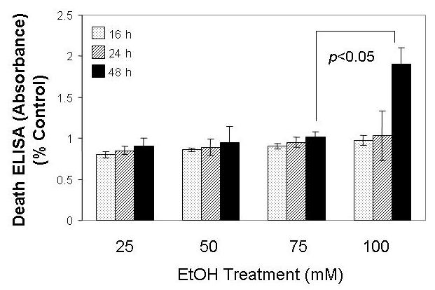
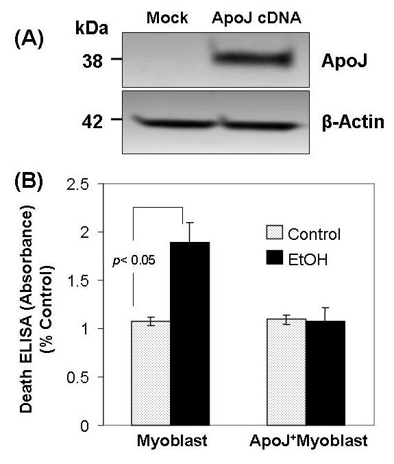
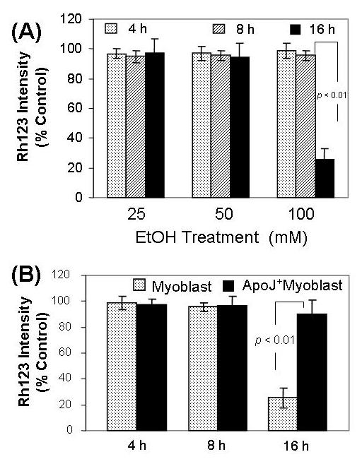
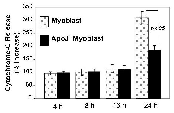
Over-consumption of ethanol (EtOH) represents a major health problem. This study was to test the cytotoxicity of EtOH in cardiac stem cells or myoblasts, and the potential protective effect of apolipoprotein-J (ApoJ), a stress-responding, chaperone-like protein in high-density lipoprotein, on EtOH-injured cardiac myoblasts. In culture, EtOH-exposed canine fetal myoblasts underwent apoptosis in a concentration- and time-dependent manner. Expression ApoJ by cDNA transfection markedly reduced EtOH-induced apoptosis in the cells. ApoJ expression also restored partially the mitochondrial membrane potential and prevented the release of cytochrome-c from mitochondria into cytoplasma. Thus, ApoJ serves as a cytoprotective protein that protects cardiac stem cells against EtOH cytotoxicity.


Figures

Fig. 1
Fig. 1
EtOH induced fetal myoblast cell apoptosis. The non-transfected fetal myoblast cells were exposed to the specified concentration of EtOH for 16 h, 24 h and 48 h, and cells were assessed for apoptosis by analyzing histone-associated DNA fragments.
Fig.2
Fig.2
Apo J expression transduced by cDNA transfection prevents EtOH - induced apoptosis in fetal myoblast cells. Fig. 2 A. Immunoblot analysis showing ApoJ expression in fetal myoblasts transfected with or without ApoJ. Total cellular proteins extracted from fetal myoblasts and from fetal myoblasts transfected with ApoJ were subjected to 10% SDS-PAGE, then transferred to polyvinylidene fluoride membranes, blotted with anti-Apo J and anti-β-actin antibody, and detected by enhanced chemiluminescence. Anti-β-actin antibody served as a control for equal loading. Fig. 2 B. Apoptosis of ApoJ – transfected or mock control canine myoblasts exposed to EtOH. Both fetal myoblasts transfected with or without ApoJ were exposed to 100 mM EtOH for 48 h, and apoptosis was analyzed for histone-associated DNA fragments.
Fig. 3
Fig. 3
ApoJ expression restores the mitochondrial membrane potential in fetal myoblasts exposed to EtOH. Fig.3A. Effect of EtOH on mitochondrial membrane potential in fetal myoblast cells. The non - transfected fetal myoblast cells were exposed to 25 mM, 50 mM and 100 mM EtOH for 4 h, 8 h and 16 h, then loaded with 1 μM rhodamine 123 (Rh123). After washing, the cells were analyzed for fluorescence at 480/530 nm (excitation/emission) in a fluorescence plate reader. Fig 3B. Effects of ApoJ on EtOH – induced mitochondrial dysfunction. Both fetal myoblasts transfected with or without ApoJ were exposed to 100 mM EtOH for 4 h, 8 h and 16 h, then loaded with 1 μM rhodamine 123 (Rh123). After washing, the cells were analyzed for fluorescence at 480/530 nm (excitation/emission) in a fluorescence plate reader.
Fig. 4
Fig. 4
Effects of EtOH on cytochrome-c release in ApoJ-transfected or mock control fetal myoblasts. Both fetal myoblasts transfected with or without ApoJ were exposed to 100 mM EtOH for 4h, 8 h, 16 h and 24 h, and the cytosolic fractions were obtained by centrifugation. Cytochrome-c levels were assayed by ELISA, with absorbance determined using a microplate reader at 450/575 nm (excitation/emission) dual wavelengths. The results were expressed as a percentage of the untreated control ± SEM of at least three plates per group in three replicate experiments.

References

    1. Di Gennaro C, Biggi A, Barilli AL, Fasoli E, Carra N, Novarini A, Delsignore R, Montanari A. Endothelial dysfunction and cardiovascular risk profile in long-term withdrawing alcoholics. J Hypertens. 2007;25:367–73. - PubMed
    1. Rubert G, Minana R, Pascual M, Guerri C. Ethanol exposure during embryogenesis decreases the radial glial progenitorpool and affects the generation of neurons and astrocytes. J Neurosci Res. 2006;84:483–96. - PubMed
    1. Kang K, Oh YK, Choue R, Kang SJ. Scutellariae radix extracts suppress ethanol-induced caspase-11 expression and cell death in N(2)a cells. Brain Res Mol Brain Res. 2005;142:139–45. - PubMed
    1. Bhave SV, Hoffman PL. Ethanol promotes apoptosis in cerebellar granule cells by inhibiting the trophic effect of NMDA. J Neurochem. 1997;68:578–86. - PubMed
    1. Li Y, King MA, Meyer EM. alpha7 nicotinic receptor-mediated protection against ethanol-induced oxidative stress and cytotoxicity in PC12 cells. Brain Res. 2000;861:165–7. - PubMed

Publication types