. 2007 May 25;282(21):15404-15.
doi: 10.1074/jbc.M701638200. Epub 2007 Apr 5.

Thrombospondin-1 inhibits nitric oxide signaling via CD36 by inhibiting myristic acid uptake

Affiliations

Thrombospondin-1 inhibits nitric oxide signaling via CD36 by inhibiting myristic acid uptake

Jeff S Isenberg et al. J Biol Chem. .

Abstract

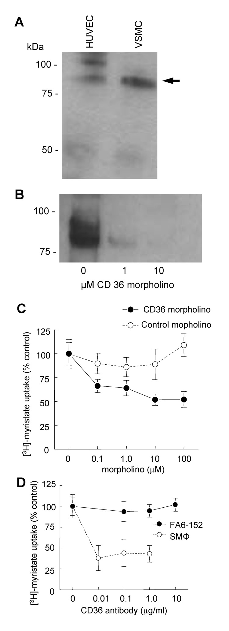
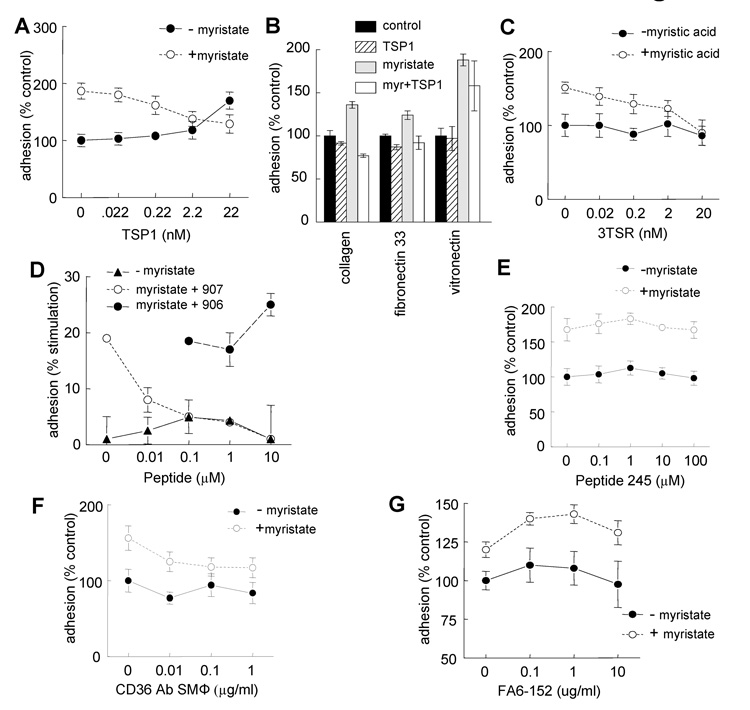
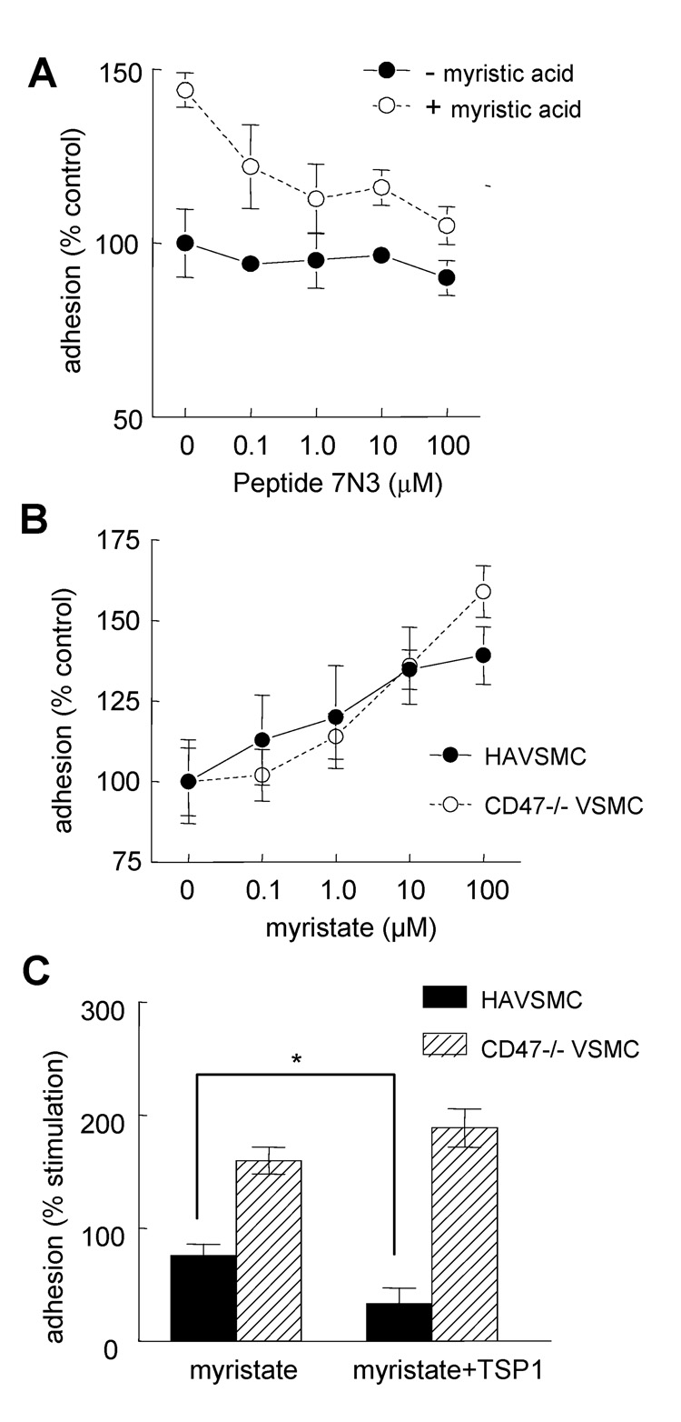
Although CD36 is generally recognized to be an inhibitory signaling receptor for thrombospondin-1 (TSP1), the molecular mechanism for transduction of this signal remains unclear. Based on evidence that myristic acid and TSP1 each modulate endothelial cell nitric oxide signaling in a CD36-dependent manner, we examined the ability of TSP1 to modulate the fatty acid translocase activity of CD36. TSP1 and a CD36 antibody that mimics the activity of TSP1 inhibited myristate uptake. Recombinant TSP1 type 1 repeats were weakly inhibitory, but an anti-angiogenic peptide derived from this domain potently inhibited myristate uptake. This peptide also inhibited membrane translocation of the myristoylated CD36 signaling target Fyn and activation of Src family kinases. Myristate uptake stimulated cGMP synthesis via endothelial nitric-oxide synthase and soluble guanylyl cyclase. CD36 ligands blocked myristate-stimulated cGMP accumulation in proportion to their ability to inhibit myristate uptake. TSP1 also inhibited myristate-stimulated cGMP synthesis by engaging its receptor CD47. Myristate stimulated endothelial and vascular smooth muscle cell adhesion on type I collagen via the NO/cGMP pathway, and CD36 ligands that inhibit myristate uptake blocked this response. Therefore, the fatty acid translocase activity of CD36 elicits proangiogenic signaling in vascular cells, and TSP1 inhibits this response by simultaneously inhibiting fatty acid uptake via CD36 and downstream cGMP signaling via CD47.


Figures

Fig. 1
Fig. 1. Myristic acid uptake into vascular cells is inhibited by exogenous TSP1 and some CD36 binding peptides
A, D–F HUVEC or C, HAVSMC (5 × 10⁴ cells/well) were plated in 24-well culture plates in EGM + 2% FCS or SM-GM + 2% FCS respectively and weaned over 48 h to EBM or SM-BM + 0.1% FAF-BSA. A, Cells were then treated with [3H]-myristic acid complexed to FAF-BSA for the indicated time intervals, and uptake into cells was determined following lysis by scintillation counting. B, [3H]-Myristic acid uptake after 5 min into HUVEC was determined in the presence of the indicated concentrations of TSP1, its type 1 repeats (3TSR), its C-terminal domain (CBD), laminin or vitronectin. C, Myristic acid uptake after 5 min into HAVSMC was determined in the presence of TSP1 (0.22 – 22 nM). D–F, Uptake into HUVEC after 5 min was determined in the presence of CD36 binding peptides p907, p245 and p906 (0.1 – 10 µM, D), the CD47 binding peptide p7N3 (1 – 100 µM, E), or the CD36-specific translocase inhibitor sulfosuccinimidyl oleate (F).
Fig. 2
Fig. 2. Morpholino suppression of CD36 and a CD36 blocking antibody decrease myristate uptake into vascular cells
A, HUVEC and human aortic VSMC were cultured in standard growth medium, surface labeled with biotin, lysed, and immunoprecipitated using a monoclonal antibody to CD36. Equal amounts of protein were electrophoresed, transferred to membranes, and detected using strepavidin-peroxidase and chemiluminescent detection. B, HAVSMC grown under standard growth conditions and treated for 48 with a CD36 morpholino (0 – 10 µM) and CD36 expression determined. C, HUVEC (5 × 10⁴ cells/well) were plated in 24-well culture plates in EGM + 2% FCS, treated with an antisense CD36 or control morpholino and weaned over 48 h to EBM + 0.1% BSA. Cells were then treated with [3H]-myristic acid complexed to FAF-BSA for 5 min., and uptake into cells was determined. D, HUVEC were plated in 24-well culture plates in EGM + 2% FCS and weaned over 48 h to EBM + 0.1% FAF-BSA. Cells were then treated an agonist CD36 monoclonal antibody clone SMΦ or antagonist antibody clone FA6-152 at the indicated concentrations, incubated with [3H]-myristic acid complexed to FAF-BSA for 5 min., and uptake into cells was determined as described.
Fig. 3
Fig. 3. Inhibition of CD36-mediated myristic acid uptake into endothelial cells by a TSP1-based peptide blocks rapid membrane translocation of Fyn and activation of Src kinases
A, HUVEC were plated into 100 mm plates and cultured in 2% FCS EGM to 80% confluence and serum starved over 24 h. The next day, cells were treated in EBM containing 0.1% FAF-BSA with or without peptide-907 (10 µM) for 15 min, ± myristate (10 µM) or EBM-1% FCS for 60 min. Cells were lysed, and the homogenate was separated by ultracentrifugation into membrane (P100) and cytosol fractions (S100). Proteins were separated by electrophoresis and analyzed by western blotting using Fyn and actin antibodies. B, HUVEC were starved overnight in 1% FBS-EBM, rinsed and treated with the indicated reagents in EBM with 0.1% FAF-BSA: p907 at 10 µM for 15 min before the addition of VEGF or Myr. VEGF was added at 20 ng/ml for 5 min and myristate at 10 µM for 15 min. Cells were then rinsed with PBS and collected in SDS sample buffer. Westerns blots were probed with anti-Src-Y416 (Cell Signaling) or anti-Src (Cell Signaling). Membranes were reprobed with anti-actin antibody for loading control. Results presented are representative of three independent experiments.
Fig. 4
Fig. 4. TSP1 prevents myristic acid-stimulated cGMP accumulation in vascular cells mediated by CD36 and NOS
A–B, HUVEC plated at 5 × 10³ cells/well were weaned over 48 h from serum and then treated in EBM containing 0.1% FAF-BSA with 0.1 – 100 µM myristic acid for 5 min (A), or with 10 µM myristic acid for the indicated times (B). Cells were lysed and cGMP levels determined via ELISA. C,D,F,G, Endothelial cells were pretreated with TSP1 for 15 min (1 µg/ml, C), an antisense CD36 or control morpholino (10 µM D) for 48 hours, SSO for 15 min (F), or L-NAME (500 µM, G) and then with 10 µM myristate/FAF-BSA for 5 min. E, HAVSMC were treated with an antisense CD36 or control morpholino (10 µM D) for 48 hours and then with 10 µM myristate/FAF-BSA for 5 min. Cells were then lysed and cGMP levels determined. Results are representative of those obtained in three independent experiments.
Fig. 5
Fig. 5. A CD36-binding peptide prevents myristate-driven cGMP accumulation in endothelial cells
HUVEC cells plated at 5 × 10³ cells/well were weaned over 48 h from serum then treated in EBM containing 0.1% FAF-BSA and pre-treated with the translocase inhibiting CD36-binding peptide (p907, A) or control (p906, B) (10 µM) for 15 min, treated with myristate (10 µM) for 5 min, and cGMP levels determined via ELISA.
Fig. 6
Fig. 6. Myristic acid stimulates endothelial cell adhesion to type I collagen via CD36/NOS/sGC/cGK signaling
HUVEC (104 cells/well) were plated in 96-well plates pre-coated with type I collagen (3 µg/ml) in EBM+0.1% FAF-BSA and the indicated concentrations of (A) myristic acid or oleic acid, (C) SSO ± myristic acid (10 µM), (D) cells pretreated for 48 h with an antisense CD36 or mismatched control morpholino (10 µM) ± myristic acid (10 µM), (E) myristic acid ± the NOS inhibitor L-NAME (500 µM), (F) the soluble guanylyl cyclase inhibitor ODQ (0.01 – 10 µM) ± myristic acid (10 µM), or (G) the cGK inhibitor Rp-8-pCPT-cGMPs (10 µM) ± myristic acid (10 µM) or DEA/NO (10 µM) and incubated for 1 h at 37° C and 5% CO2. (B) HUVEC (104 cell/well) were plated in 96-well plates pre-coated with type I collagen (3 µg/ml), vitronectin (2 µg/ml), fibronectin (3 µg/ml), or FN-33 (3 µg/ml) in EBM + 0.1% FAF-BSA ± myristic acid (10 µM). Adherent cells were quantified after staining with crystal violet at 570 nm. Results are expressed as the mean ± SD of triplicates and are representative of at least three experiments.
Fig. 7
Fig. 7. CD36 ligands that inhibit fatty acid translocation prevent myristate-stimulated cell adhesion
HUVEC (104 cell/well) were plated in 96-well plates pre-coated with type I collagen (3 µg/ml) in EBM + 0.1% FAF-BSA ± myristic acid (10 µM) and the indicated concentrations of (A) TSP1, (C) 3TSR, (D) CD36-binding peptide 907 and control peptide 906, (E) noninhibitory CD36-binding peptide 245, (F) the uptake-blocking CD36 antibody SMΦ, or (G) the nonblocking CD36 antibody FA6-152 and incubated for 1 h at 37° C and 5% CO2. (B) HAVSMC (104 cell/well) were plated in 96-well plates pre-coated with type I collagen (3 µg/ml), vitronectin (1 µg/ml), or FN-33 (1.5 µg/ml) in SM-BM + 0.1% FAF-BSA ± myristic acid (10 µM) ± TSP1 (2.2 nM). Adhesion was quantified by colorimetric assay at 570 nm. Results are expressed as the mean ± SD of triplicates and are representative of at least three experiments.
Fig. 8
Fig. 8. CD47 is not necessary for stimulation of adhesion by myristate but is required for inhibition of this response by low concentrations of TSP1
HUVEC (104 cell/well) were plated in 96-well plates pre-coated with type I collagen (3 µg/ml) in EBM + 0.1% FAF-BSA and the indicated concentrations of (A) the CD47-binding peptide p7N3 (0.1 – 100 µM) ± myristic acid (10 µM) and incubated for 1 h at 37° C and 5% CO2. Adhesion was quantified by colorimetric assay at 570 nm. (B), HAVSMC and CD47 null VSMC (104 cell/well) were plated in 96-well plates pre-coated with type I collagen (3 µg/ml) in SM-BM + 0.1% FAF-BSA and the indicated concentrations of myristic acid, or (C) myristic acid (10 µM) ± TSP1 (2.2 nM) and adhesion determined by colorimetric assay at 570 nm. Results are expressed as the mean ± SD of triplicates and representative of at least three experiments.
Fig. 9
Fig. 9. Model for CD36-dependent activities of TSP1 and other CD36 ligands
Uptake of myristic acid via the fatty acid translocase (FAT) activity of CD36 is potently inhibited by the TSP1-derived mimetic p907 and by the angiogenesis-inhibiting CD36 antibody SMΦ (10). TSP1 inhibits FAT activity, and CD36 antibody FA6-152 is known to antagonize the anti-angiogenic activity of TSP1 (10,12,13) but does not itself inhibit FAT. In serum starved endothelial cells, inhibiting myristic acid transport is proposed to limit precursor for synthesis of myristoyl-CoA via the acyl-CoA synthase (ACSL3), which is then transferred by N-myristoyl transferases (NMT) to proteins bearing subterminal Gly residues exposed by Met aminopeptidases (MetAP). Of these, MetAP2 is the known target of several angiogenesis inhibitors (58), suggesting convergence of the signaling pathways induced by these inhibitors and TSP1. Limiting myristoylation prevents translocation of Fyn and other myristoylated proteins to membranes and indirectly limits eNOS activity. TSP1 at 1–10 nM limits cGMP signaling by inhibiting myristate uptake via CD36 and at 10–100 pM also inhibits sGC activation via CD47 (22).

References

    1. Carmeliet P. Nature. 2005;438(7070):932–936. - PubMed
    1. Lawler J, Detmar M. Int J Biochem Cell Biol. 2004;36(6):1038–1045. - PubMed
    1. Nyberg P, Xie L, Kalluri R. Cancer Res. 2005;65(10):3967–3979. - PubMed
    1. Calzada MJ, Zhou L, Sipes JM, Zhang J, Krutzsch HC, Iruela-Arispe ML, Annis DS, Mosher DF, Roberts DD. Circ Res. 2004;94(4):462–470. - PubMed
    1. Chandrasekaran L, He CZ, Al-Barazi H, Krutzsch HC, Iruela-Arispe ML, Roberts DD. Mol Biol Cell. 2000;11(9):2885–2900. - PMC - PubMed

Publication types

MeSH terms