

Substrate chemistry-dependent conformations of single laminin molecules on polymer surfaces are revealed by the phase signal of atomic force microscopy
- PMID: 17416620
- PMCID: PMC1914422
- DOI: 10.1529/biophysj.106.102491
Substrate chemistry-dependent conformations of single laminin molecules on polymer surfaces are revealed by the phase signal of atomic force microscopy
Abstract
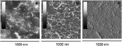
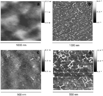
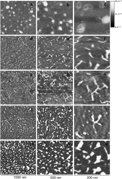
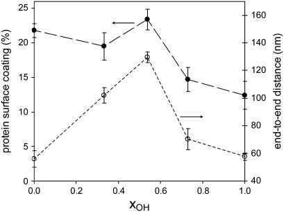
The conformation of single laminin molecules adsorbed on synthetic substrates is directly observed making use of the phase magnitude in tapping mode atomic force microscopy (AFM). With AFM, it is not possible to differentiate the proteins on the substrate if use is made of the height signal, since the roughness of the material becomes of the same order of magnitude as the adsorbed protein, typically 10 nm height. This work shows how AFM can be exploited to reveal protein conformation on polymer materials. Different laminin morphologies are observed on a series of different copolymers based on ethyl acrylate and hydroxyethyl acrylate as a function of the surface density of -OH groups: from globular to completely extended morphologies of the protein molecules are obtained, and the onset of laminin network formation on some substrates can be clearly identified. The results stress the importance of the underlying synthetic substrate's surface chemistry for the biofunctional conformation of adsorbed proteins.
Figures
References
-
- Boyan, B. D., T. W. Hummert, D. D. Dean, and Z. Schwartz. 1996. Role of material surfaces in regulating bone and cartilage cell response. Biomaterials. 17:137–146. - PubMed
-
- Anselme, K. 2000. Osteoblast adhesion on biomaterials. Biomaterials. 21:667–681. - PubMed
-
- Alberts, B., A. Johnson, J. Lewis, M. Raff, K. Roberts, and P. Walter. 2004. Molecular Biology of the Cell. Garland Science, NY.
-
- Hynes, R. O. 2002. Integrins: bidirectional, allosteric signaling machines. Cell. 110:673–687. - PubMed
-
- Yamada, K. M. 1991. Adhesive recognition sequences. J. Biol. Chem. 266:12809–12812. - PubMed
Publication types
MeSH terms
Substances
LinkOut - more resources
Full Text Sources
Other Literature Sources
Miscellaneous
