. 2007 Jul 1;93(1):103-12.
doi: 10.1529/biophysj.106.100370. Epub 2007 Apr 6.

Pore formation by a Bax-derived peptide: effect on the line tension of the membrane probed by AFM

Affiliations

Pore formation by a Bax-derived peptide: effect on the line tension of the membrane probed by AFM

Ana J García-Sáez et al. Biophys J. .

Abstract

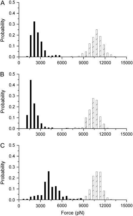
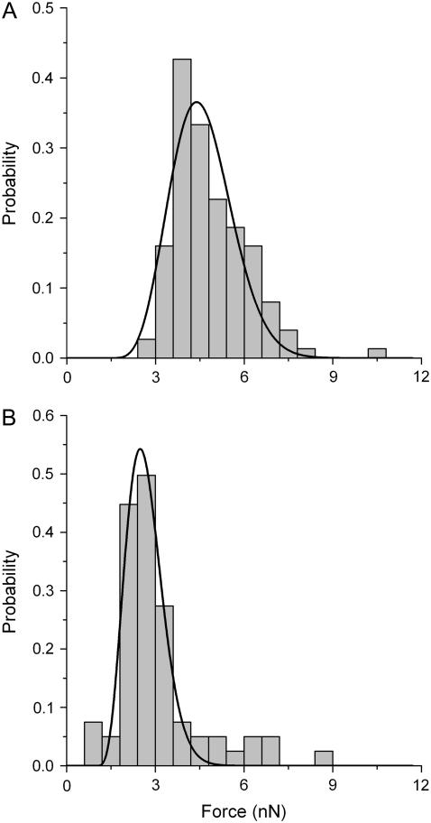
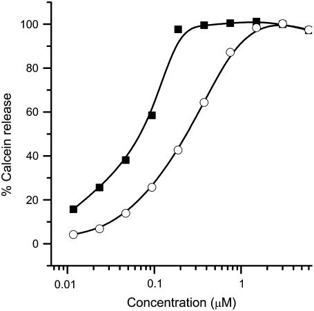
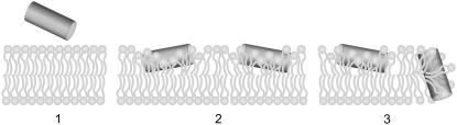
Bax is a critical regulator of physiological cell death that increases the permeability of the outer mitochondrial membrane and facilitates the release of the so-called apoptotic factors during apoptosis. The molecular mechanism of action is unknown, but it probably involves the formation of partially lipidic pores induced by Bax. To investigate the interaction of Bax with lipid membranes and the physical changes underlying the formation of Bax pores, we used an active peptide derived from helix 5 of this protein (Bax-alpha5) that is able to induce Bax-like pores in lipid bilayers. We report the decrease of line tension due to peptide binding both at the domain interface in phase-separated lipid bilayers and at the pore edge in atomic force microscopy film-rupture experiments. Such a decrease in line tension may be a general strategy of pore-forming peptides and proteins, as it affects the energetics of the pore and stabilizes the open state.


Figures

FIGURE 1
FIGURE 1
Kinetics of the morphological changes induced by Bax-α5 in domain-exhibiting membranes. Bax-α5 was added to DOPC/SM/Chol (1:1:0.67) supported bilayers containing 0.01% DiD to L/P = 109 25 s after the film starts. Confocal images were taken at 25-s intervals for 10 min. The selected times are shown.
FIGURE 2
FIGURE 2
AFM topographical images of supported lipid bilayer composed of DOPC/SM/Chol (1:1:0.67) and imaged at 25°C. Bright and dark regions represent SM-rich and DOPC-rich phases, respectively. (A) AFM topographical appearance of a freshly prepared supported membrane at equilibrium. (B) The same supported bilayer as in A, but 1 h after addition of Bcl-xL-α5 at L/P = 14 molar ratio. (C) Surface of a supported bilayer with the same lipid composition as in A, but 30 min after addition of Bax-α5 at L/P = 109 molar ratio.
FIGURE 3
FIGURE 3
Typical approach part of a force curve measured on the Lo domain in a DOPC/SM/Chol (1:1:0.67) supported bilayer on mica. The force applied by the cantilever is represented as a function of the z-piezo displacement. The discontinuity in the curve corresponds to the yield force required for the AFM tip to punch through the bilayer (arrow).
FIGURE 4
FIGURE 4
Histograms of yield forces measured on a DOPC/SM/Chol (1:1:0.67) bilayer at different Bax-α5 concentrations: (A) in the absence of peptide; (B) at L/P = 109; (C) at L/P = 55; (D) at L/P = 27; and (E) at L/P = 14. The black bars correspond to the Ld phase, whereas the dashed bars correspond to the Lo phase. The histograms are normalized for better comparison (the number of points varied between 70 and 350).
FIGURE 5
FIGURE 5
Histograms of yield forces measured on a DOPC/SM/Chol (1:1:0.67) bilayer at different Bcl-xL-α5 concentrations: (A) in the absence of peptide; (B) at L/P =55; and (C) at L/P = 14. The black bars correspond to the Ld phase, whereas the dashed bars correspond to the Lo phase. The histograms are normalized for better comparison (the number of points varied between 70 and 350).
FIGURE 6
FIGURE 6
Normalized histograms of yield forces measured on a POPC supported bilayer (A) in the absence and (B) at a ratio of L/P ≈ 5000 of Bax-α5. The solid lines correspond to the fitted function of probability density from Eq. 14. The number of points was 66 in A and 124 in B.
FIGURE 7
FIGURE 7
Permeabilizing activity of Bax-α5 in LUVs. The percentage of calcein release, according to Eq. 2, is represented as a function of the concentration of added peptide. Vesicles were composed of egg PC (solid squares) or PC/SM/Chol (1:1:1) (open circles).
FIGURE 8
FIGURE 8
Proposed mechanism of pore formation by Bax-α5. Bax-α5 is an amphipathic molecule with hydrophobic residues on one side (blue) and net positive charge on the other side (red). (1) Electrostatic forces favor the interaction with the polar surface of the membrane. (2) Insertion of the peptide at the interface between the headgroup region and the hydrophobic region provokes lipid distortion and induces stress in the bilayer. (3) After a threshold concentration, Bax-α5 forms pores of a toroidal nature and stabilizes them by reducing the line tension at the pore rim.

References

    1. Adams, J. M., and S. Cory. 1998. The Bcl-2 protein family: arbiters of cell survival. Science. 281:1322–1326. - PubMed
    1. Newmeyer, D. D., and S. Ferguson-Miller. 2003. Mitochondria: releasing power for life and unleashing the machineries of death. Cell. 112:481–490. - PubMed
    1. Wei, M. C., W. X. Zong, E. H. Cheng, T. Lindsten, V. Panoutsakopoulou, A. J. Ross, K. A. Roth, G. R. MacGregor, C. B. Thompson, and S. J. Korsmeyer. 2001. Proapoptotic BAX and BAK: a requisite gateway to mitochondrial dysfunction and death. Science. 292:727–730. - PMC - PubMed
    1. Zong, W. X., T. Lindsten, A. J. Ross, G. R. MacGregor, and C. B. Thompson. 2001. BH3-only proteins that bind pro-survival Bcl-2 family members fail to induce apoptosis in the absence of Bax and Bak. Genes Dev. 15:1481–1486. - PMC - PubMed
    1. Crompton, M. 2000. Bax, Bid and the permeabilization of the mitochondrial outer membrane in apoptosis. Curr. Opin. Cell Biol. 12:414–419. - PubMed

Publication types