

Structures of tRNAs with an expanded anticodon loop in the decoding center of the 30S ribosomal subunit
- PMID: 17416634
- PMCID: PMC1869038
- DOI: 10.1261/rna.367307
Structures of tRNAs with an expanded anticodon loop in the decoding center of the 30S ribosomal subunit
Abstract
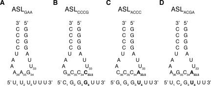
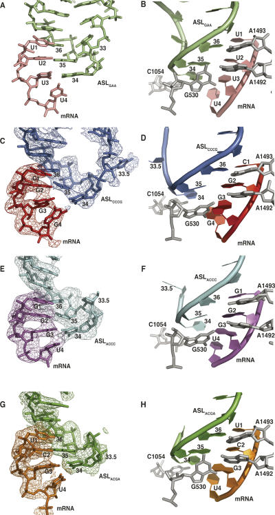
During translation, some +1 frameshift mRNA sites are decoded by frameshift suppressor tRNAs that contain an extra base in their anticodon loops. Similarly engineered tRNAs have been used to insert nonnatural amino acids into proteins. Here, we report crystal structures of two anticodon stem-loops (ASLs) from tRNAs known to facilitate +1 frameshifting bound to the 30S ribosomal subunit with their cognate mRNAs. ASL(CCCG) and ASL(ACCC) (5'-3' nomenclature) form unpredicted anticodon-codon interactions where the anticodon base 34 at the wobble position contacts either the fourth codon base or the third and fourth codon bases. In addition, we report the structure of ASL(ACGA) bound to the 30S ribosomal subunit with its cognate mRNA. The tRNA containing this ASL was previously shown to be unable to facilitate +1 frameshifting in competition with normal tRNAs (Hohsaka et al. 2001), and interestingly, it displays a normal anticodon-codon interaction. These structures show that the expanded anticodon loop of +1 frameshift promoting tRNAs are flexible enough to adopt conformations that allow three bases of the anticodon to span four bases of the mRNA. Therefore it appears that normal triplet pairing is not an absolute constraint of the decoding center.
Figures
References
-
- Brünger, A.T., Adams, P.D., Clove, G.M., Delano, W.L., Gros, P., Grosse-Kunstleve, R.W., Jiang, J.-S., Kuszewski, J., Nilges, M., Pannu, N.S., et al. Crystallography and NMR system: A new software suite for macromolecular structure determination. Acta Crystallogr. D Biol. Crystallogr. 1998;54:905–921. - PubMed
-
- Carter, A.P., Clemons, W.M., Brodersen, D.E., Morgan-Warren, R.J., Wimberly, B.T., Ramakrishnan, V. Functional insights from the structure of the 30S ribosomal subunit and its interactions with antibiotics. Nature. 2000;407:340–348. - PubMed
-
- CCP4. The CCP4 suite: Programs for protein crystallography. Acta Crystallogr. D Biol. Crystallogr. 1994;50:760–763. - PubMed
-
- Clemons W.M., Jr, Brodersen, D.E., McCutcheon, J.P., May, J.L., Carter, A.P., Morgan-Warren, R.J., Wimberly, B.T., Ramakrishnan, V. Crystal structure of the 30S ribosomal subunit from Thermus thermophilus: Purification, crystallization, and structure determination. J. Mol. Biol. 2001;310:827–843. - PubMed
Publication types
MeSH terms
Substances
Grants and funding
LinkOut - more resources
Full Text Sources