. 2007 Jun;13(6):890-8.
doi: 10.1261/rna.359007. Epub 2007 Apr 6.

Induction of drug resistance and transformation in human cancer cells by the noncoding RNA CUDR

Affiliations

Induction of drug resistance and transformation in human cancer cells by the noncoding RNA CUDR

Wing Pui Tsang et al. RNA. 2007 Jun.

Abstract

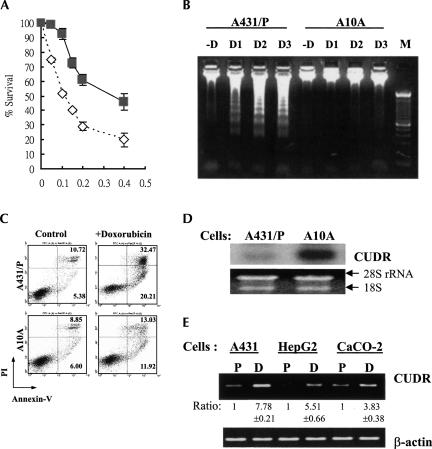
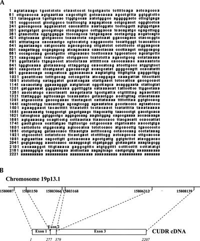
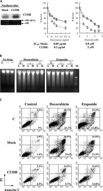
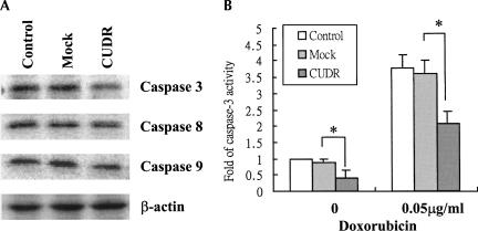
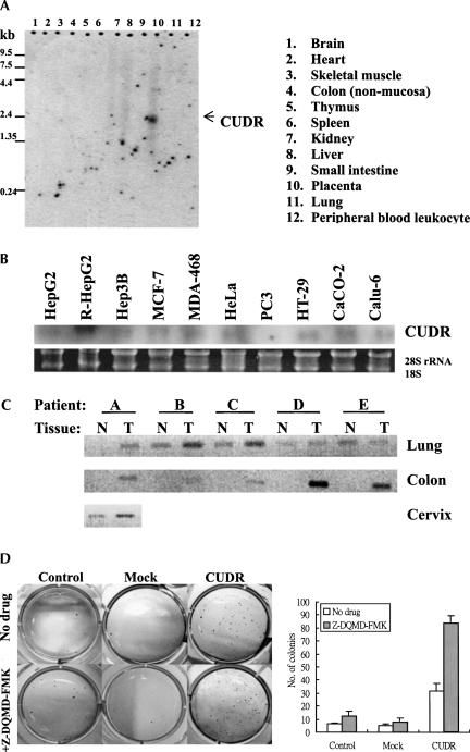
Refractory to apoptosis induced by anticancer drugs is one of the major causes of drug resistance in human cancers. The involvement of noncoding RNA (ncRNA) in cancer cell drug resistance has not yet been reported. By using the technique of RT-PCR-based differential display, a novel gene, cancer up-regulated drug resistant (CUDR) gene, was found to be overexpressed in a doxorubicin-resistant subline of human squamous carcinoma A431 and A10A cells, which were also more resistant to drug-induced apoptosis. The full-length CUDR mRNA transcript is approximately 2.2 kb as detected by Northern blot analysis and has no sequence homology with other genes identified so far. Interestingly, no distinct open reading frame was found throughout the CUDR cDNA sequence, and no recombinant protein was detected from in vitro translation or from a protein lysate of human cancer cells after CUDR transfection. Therefore, CUDR is likely to exert its function as a noncoding RNA. Stable transfection with the CUDR gene was found to induce resistance to doxorubicin and etoposide as well as drug-induced apoptosis in A431 cells. By Western blot analysis, down-regulations of caspase 3 were observed in CUDR transfectants. On the other hand, overexpression of CUDR promoted anchorage-independent growth in A431 cells. Results from the present study suggest that CUDR may likely regulate the drug sensitivity and promote cellular transformation at least through caspase 3-dependent apoptosis.


Figures

FIGURE 1.
FIGURE 1.
The expression of CUDR, doxorubicin sensitivity, and doxorubicin-induced cell death in A431 cells. (A) The sensitivity to doxorubicin of A431 parent cells and A10A cells as detected by MTT assay. Cells were exposed to different concentrations of doxorubicin for 48 h. A431 parent cells (open diamond); A10A cells (gray square). Results were the mean value from four independent experiments, and error bars show 95% confidence intervals. (B) Doxorubicin-induced DNA fragmentation in A431 cells. The cells were exposed to 0.025 μg/mL (D1), 0.05 μg/mL (D2), or 0.1 μg/mL (D3) doxorubicin for 48 h. Forty micrograms of genomic DNA were resolved in a 1.5% agarose gel. (M) 100-bp DNA marker. (C) Flow cytometric analysis of cells staining with annexin V/PI. The cells were treated with 0.05 μg/mL doxorubicin for 48 h and then analyzed for early apoptotic cells (bottom right quadrant) and late apoptotic or dead cells (top right quadrant); the percentages of cells in the two quadrants are shown. (D) Northern blot analysis of CUDR in A431 parent and A10A cells. The 18S and 28S rRNA demonstrated equal loading of total RNA in each lane. (E) CUDR expression in the parent (P) and the doxorubicin-selected subline (D) of A431, HepG2, and CaCO-2 cells by semiquantitative RT-PCR. The signal of each band was quantitated by Kodak Digital Science 1D image analysis software version 3.0 (Eastman Kodak). The relative expression of each band was calculated with respect to the expression of the parent cells (designated as 1). Data shown are the mean value of three independent experiments. The CUDR expression in the drug-resistant cells is significantly different from that of the respective parent cells (P < 0.05).
FIGURE 2.
FIGURE 2.
Sequence analysis of CUDR cDNA. The CUDR gene was mapped to human chromosome 19p13.1 and transcribed into mRNA transcript with three exons.
FIGURE 3.
FIGURE 3.
CUDR overexpression-induced resistance to apoptotic cell death in cells upon drug treatment. (A) Stable expression of CUDR in A431 cells. The CUDR expression in mock and CUDR/pcDNA transfectants was assessed by Northern blot analysis. Sensitivity of A431 stable transfectants to doxorubicin and etoposide as detected by MTT assay. Cells were exposed to various concentrations of drugs for 48 h. Mock (empty vector) transfection (open diamond); CUDR/pcDNA transfection (gray square). Results show the mean value from four independent experiments, and error bars show 95% confidence intervals. (B) Drug-induced apoptosis in A431 cells as detected by a DNA fragmentation assay. The parent (C), mock (−), and CUDR (+) transfectants were exposed to doxorubicin ([L] 0.025 μg/mL, [H] 0.05 μg/mL) or etoposide ([L] 1 μM, [H] 2.5 μM) for 48 h. (M) 100-bp DNA marker. (C) Doxorubicin- and etoposide-induced apoptotsis in A431 cells assessed by annexin V-GFP/propidium iodide staining followed by flow cytometric analysis. Cells were stained with annexin V-GFP/PI after drug treatments for 48 h, and then analyzed for early apoptotic cells (bottom right quadrant) and late apoptotic or dead cells (top right quadrant). The percentages of cells in the two quadrants are shown.
FIGURE 4.
FIGURE 4.
(A) The basal protein levels of caspase 3, caspase 8, and caspase 9 in A431 cells. Twenty-five micrograms of total protein lysate were resolved in 15% SDS-PAGE and immunoblotted with the corresponding antibody by Western blot analysis. Experiments have been repeated at least three times, and the representative one is shown. (B) Effect of doxorubicin on caspase 3 activity in A431 cells. After treatment with 0.05 μg/mL doxorubicin for 48 h, the cells were trypsinized and lysed. Protein lysate was then subjected to a caspase 3 activity assay. Results show the mean value from four independent experiments, and error bars show 95% confidence intervals. (*) P < 0.05.
FIGURE 5.
FIGURE 5.
CUDR expression in human normal tissues and cancer tissues. (A) CUDR mRNA expression in different human normal tissues as detected by Northern blot analysis using the pre-made 12-lane human tissue Northern blot (Clontech). (B) CUDR expression in human cancer cell lines as detected by Northern blot analysis. (C) CUDR expression in cDNA pairs of matched normal (N) and tumor (T) tissues (Clontech) as detected by PCR. PCR was performed with a pair of CUDR-specific primers and then resolved in a 1.2% agarose gel. (A–E) Different patients designed by the manufacturer. The level of β-actin in each sample was also measured and did not demonstrate deviation among the samples (data not shown). (D) The effect of CUDR transfection on anchorage-independent cell growth of A431 cells with or without 2 μM caspase 3 inhibitor Z-DQMD-FMK as examined by soft agar colony formation assay. The number of colonies of each cell line is shown. Results show the mean value from three independent experiments, and error bars show 95% confidence intervals.

Similar articles

Cited by

References

    1. Cole, S.P., Deeley, R.G. Multidrug resistance mediated by the ATP-binding cassette transporter protein MRP. Bioessays. 1995;20:931–940. - PubMed
    1. Devarajan, E., Sahin, A.A., Chen, J.S., Krishnamurthy, R.R., Aggarwal, N., Brun, A.M., Sapino, A., Zhang, F., Sharma, D., Yang, X.H., et al. Down-regulation of caspase 3 in breast cancer: A possible mechanism for chemoresistance. Oncogene. 2002;21:8843–8851. - PubMed
    1. Eddy, S.R. Noncoding RNA genes and the modern RNA world. For. Genet. 2001;2:919–929. - PubMed
    1. Erdmann, V.A., Barciszewska, M.Z., Hochberg, A., de Groot, N., Barciszewski, J. Regulatory RNAs. Cell. Mol. Life Sci. 2001;58:960–977. - PMC - PubMed
    1. Fire, A., Xu, S., Montomery, M.K., Kostas, S.A., Driver, S.E., Mello, C.C. Potent and specific genetic interference by double-stranded RNA in Caenorhabditis elegans . Nature. 1998;391:806–811. - PubMed

Publication types