. 2007 Jun;189(12):4473-84.
doi: 10.1128/JB.00227-07. Epub 2007 Apr 6.

Distribution of protein A on the surface of Staphylococcus aureus

Affiliations

Distribution of protein A on the surface of Staphylococcus aureus

Andrea C DeDent et al. J Bacteriol. 2007 Jun.

Abstract

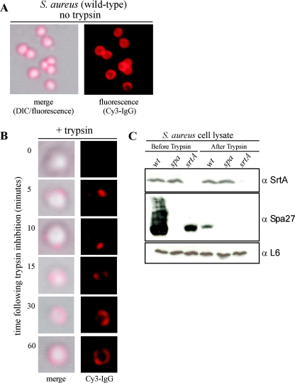
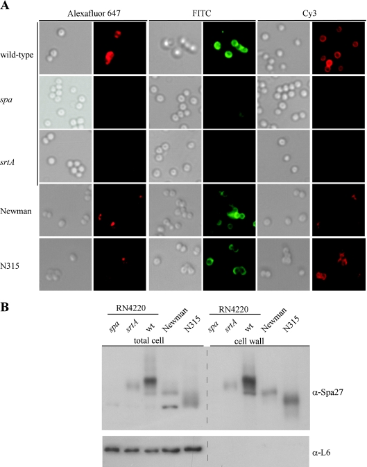
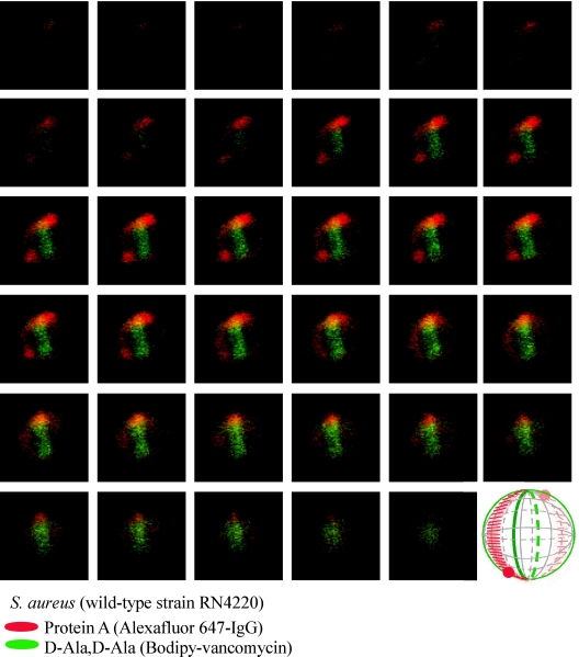
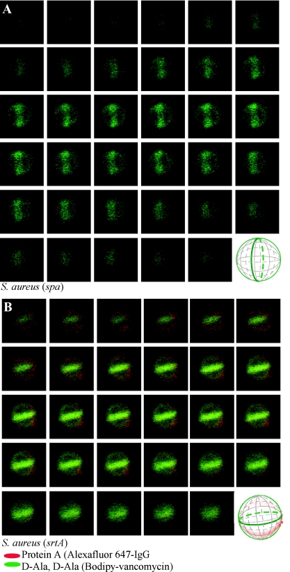
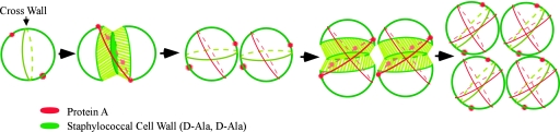
Surface proteins of Staphylococcus aureus fulfill many important roles during the pathogenesis of human infections and are anchored to the cell wall envelope by sortases. Although the chemical linkage of proteins to cell wall cross bridges is known, the mechanisms whereby polypeptides are distributed on the staphylococcal surface have not been revealed. We show here that protein A, the ligand of immunoglobulin, is unevenly distributed over the staphylococcal surface. Upon removal with trypsin, newly synthesized polypeptide is deposited at two to four discrete foci. During subsequent growth, protein A appears to be slowly distributed from these sites. When viewed through multiple focal planes by laser scanning microscopy, protein A foci are arranged in a circle surrounding the bacterial cell. This pattern of distribution requires the LPXTG sorting signal of protein A as well as sortase A, the transpeptidase that anchors polypeptides to cell wall cross bridges. A model is presented whereby protein A deposition at discrete sites coupled with cell wall synthesis enables distribution of protein A on the staphylococcal surface.


Figures

FIG. 1.
FIG. 1.
Deposition of newly synthesized protein A on the staphylococcal surface. (A) Protein A on the surface of S. aureus RN4220 cells was stained with Cy3-IgG, and DIC or fluorescence images were captured with an Olympus AX-70 fluorescence microscope and Himamatsu CCD camera. (B) Staphylococci were treated with trypsin to remove surface proteins, and the deposition of newly synthesized protein A on the cell surface at indicated times was visualized by staining with Cy3-IgG. DIC and fluorescence images were captured with a CCD camera. (C) Staphylococcal lysates before and after trypsin treatment were precipitated with trichloroacetic acid, washed in acetone, and separated on SDS-PAGE. Samples were subjected to immunoblotting with antibodies against sortase A (α-SrtA), protein A (α-Spa27), or ribosomal protein L6 (α-L6).
FIG. 2.
FIG. 2.
Distribution of protein A on the surface of staphylococcal strains. (A) S. aureus RN4220 (wild type) and isogenic spa and srtA mutants were incubated with Alexa Fluor 647-IgG, FITC-IgG, or Cy3-IgG, and DIC or fluorescence images were captured with a Olympus AX-70 fluorescence microscope and Himamatsu CCD camera. As a control for protein A distribution on human clinical isolates, S. aureus Newman and N315 strains were analyzed with the same technology. (B) Total cell extracts or cell wall fractions generated by degradation of murein sacculi with lysostaphin obtained from S. aureus RN4220 (wild-type [wt]) and isogenic spa and srtA mutants or from strains Newman and N315. Samples were separated by SDS-PAGE and then analyzed by immunoblotting with monoclonal antibody specific for protein A (α-Spa27) or with polyclonal antiserum raised against purified L6 ribosomal protein.
FIG. 3.
FIG. 3.
Protein A localization in a dividing S. aureus RN4220 cell. Laser scanning confocal microscopy images (z series with 50-nm increments) of S. aureus RN4220. Protein A was labeled with Alexa Fluor 647-IgG (red fluorescence), and cell wall pentapeptide was stained with BODIPY-vancomycin (green fluorescence). Central regions of intense green fluorescence reveal the cross wall, the cell wall layer separating two daughter cells. Each display item is derived from merged images of confocal scans of separate laser line channels. Aggregate data were used to build a three-dimensional model of protein A deposition in the cell wall, which is shown as a diagram in the lower right corner.
FIG. 4.
FIG. 4.
Protein A localization in a dividing S. aureus Newman cell. Laser scanning confocal microscopy images (z series with 50-nm increments) of S. aureus Newman. Protein A was labeled with Alexa Fluor 647-IgG (red fluorescence), and cell wall pentapeptide was stained with BODIPY-vancomycin (green fluorescence). Protein A and cell wall images of pentapeptide precursors were collected as described in the legend of Fig. 3. Aggregate data were used to build a three-dimensional model of protein A deposition in the cell wall, which is shown as a diagram in the lower right corner.
FIG. 5.
FIG. 5.
Protein A distribution in dividing S. aureus cells requires sortase A. Laser scanning confocal microscopy images (z series with 50-nm increments) of S. aureus mutants lacking the protein A gene (spa) (A) or the sortase A gene (srtA) (B). Protein A was labeled with Alexa Fluor 647-IgG (red fluorescence), and cell wall pentapeptide was stained with BODIPY-vancomycin (green fluorescence). Protein A and cell wall images of pentapeptide precursors were collected as described in the legend of Fig. 3. Aggregate data were used to build three-dimensional models of protein A deposition in the cell wall, which are shown as diagrams in the lower right corners of the panels.
FIG. 6.
FIG. 6.
Protein A distribution in dividing S. aureus cells requires a functional LPXTG sorting signal. S. aureus mutants lacking the protein A gene (spa) were transformed with plasmid encoding wild-type protein A (Spa) (A) or a variant lacking the LPXTG motif (SpaΔLPXTG) (B). Laser scanning confocal microscopy images (z series with 50-nm increments) of S. aureus mutants lacking the protein A gene (spa) or the sortase A gene (srtA). Protein A and cell wall images of pentapeptide precursors were collected as described in the legend of Fig. 3. Aggregate data were used to build three-dimensional models of protein A deposition in the cell wall, which are shown as diagrams in the lower right corners of the panels.
FIG. 7.
FIG. 7.
Diagram to illustrate a hypothesis on the deposition of protein A in the cell wall envelope of S. aureus. The cross wall, a thick layer of newly synthesized peptidoglycan, separates two newly formed daughter cells and is indicated as a green ring structure. Two to four foci of protein A staining (red dots) mark sites for the secretion and deposition of newly synthesized surface protein into murein sacculi. As cells grow and the cross wall expands, surface protein deposition forms a ring-like structure that traverses areas of cell wall synthesis. Once cell wall synthesis and separation as well as surface protein ring formation have been completed, new cross walls and foci of protein A deposition are formed perpendicular to previous planes of division and surface protein anchoring.

References

    1. Bae, T., and O. Schneewind. 2003. The YSIRK-G/S motif of staphylococcal protein A and its role in efficiency of signal peptide processing. J. Bacteriol. 185:2910-2919. - PMC - PubMed
    1. Barnett, T. C., and J. R. Scott. 2002. Differential recognition of surface proteins in Streptococcus pyogenes by two sortase gene homologs. J. Bacteriol. 184:2181-2191. - PMC - PubMed
    1. Boneca, I. G., Z. H. Huang, D. A. Gage, and A. Tomasz. 2000. Characterization of Staphylococcus aureus cell wall glycan strands, evidence for a new β-N-acetylglucosaminidase activity. J. Biol. Chem. 275:9910-9918. - PubMed
    1. Carlsson, F., M. Stalhammar-Carlemalm, K. Flardh, C. Sandin, E. Carlemalm, and G. Lindahl. 2006. Signal sequence directs localized secretion of bacterial surface proteins. Nature 442:943-946. - PubMed
    1. Cole, R. M. 1965. Bacterial cell wall replication followed by immunofluorescence. Bacteriol. Rev. 29:326-344. - PMC - PubMed

Publication types

MeSH terms