. 2007 Jun;189(11):4153-60.
doi: 10.1128/JB.01909-06. Epub 2007 Apr 6.

Purification and characterization of mycobacterial phospholipase A: an activity associated with mycobacterial cutinase

Affiliations

Purification and characterization of mycobacterial phospholipase A: an activity associated with mycobacterial cutinase

Sarah K Parker et al. J Bacteriol. 2007 Jun.

Abstract

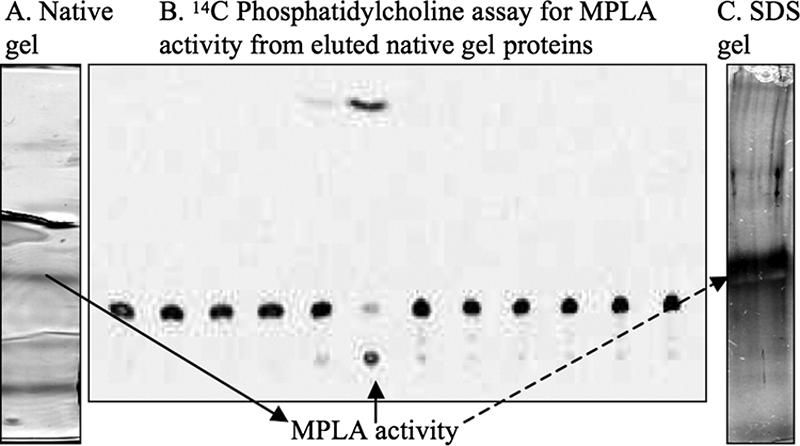
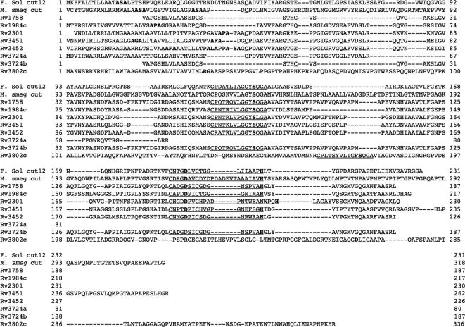
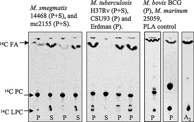
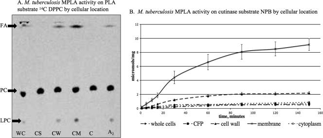
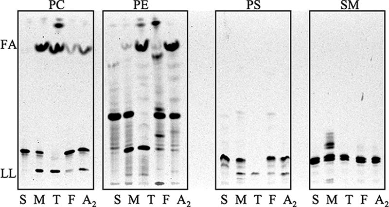
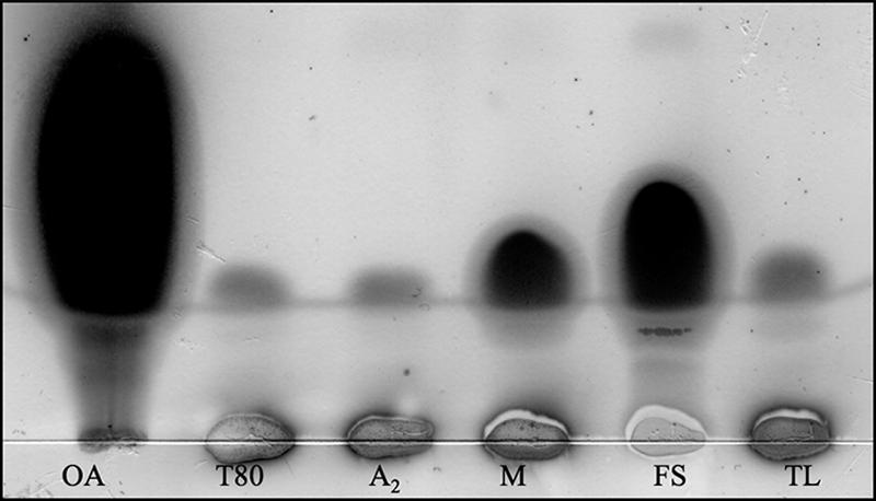
We describe mycobacterial phospholipase A activity (MPLA) and, using reverse genetics, have associated this activity with putative mycobacterial cutinase. PLAs, which hydrolyze fatty acids on phospholipids, play a significant role in human inflammatory states and disease pathogenesis. In prokaryotes, the recognition of their role in virulence is more recent. Cutinases are serine esterases whose primary substrate is cutin, the waxy exterior layer of plants. Mycobacterium tuberculosis has maintained seven putative cutinases, though it should not encounter cutin; we demonstrate that known cutinases and MPLA cleave phospholipids in a PLA-type manner and also hydrolyze Tween. We analyzed cutinase motifs in mycobacteria and found the motif very prevalent. All mycobacteria tested had MPLA activity. These studies suggest an alternative use for putative cutinases by the M. tuberculosis group that is likely related to MPLA activity and lipid metabolism.


Figures

FIG. 1.
FIG. 1.
(A) Native gel after column purification of M. smegmatis MPLA activity. (B) The gel was cut into 12 fractions, protein was eluted, and radiolabel 14C TLC was performed for MPLA activity; activity is in the sixth fraction from the indicated band (solid arrow) on the native gel. Mass spectrometry was done on this fraction. (C) An SDS gel of the active fraction showed one band at 30 kDa (dashed arrow).
FIG. 2.
FIG. 2.
Alignment of a known cutinase, F. solani cut12, with M. smegmatis cutinase that we found through reverse genetics of MPLA activity and the eight putative H37Rv cutinases. Active sites (S, D, and H) are in bold; the cutinase motif is underlined. All putative cutinases except 3724a have a cutinase motif, and all except 1758 and 3724b appear to have secretion signals.
FIG. 3.
FIG. 3.
MPLA activity determined by TLC of various mycobacterial strains. Cell pellets (P) and culture supernatants (S) were assayed using [14C]dipalmitoyl PC labeled on both palmitic acids. The presence of [14C]FA and [14C]LPC indicates hydrolysis of [14C]dipalmitoyl PC from MPLA activity. A2, snake venom PLA2. Culture supernatant from H37Rv is lyophilized culture filtrate (shown). Nonlyophilized H37Rv culture supernatant and nonlyophilized ammonium sulfate-precipitated H37Rv culture supernatant were also tested and were inactive; M. marinum 11566 (P+S) and M. marinum 25059 (P) were tested and were active (data not shown).
FIG. 4.
FIG. 4.
(A) TLC of M. tuberculosis H37Rv MPLA activity by cellular location using [14C]dipalmitoyl PC (DPPC) labeled on both palmitic acids. The presence of [14C]FA and [14C]LPC indicates hydrolysis of [14C]dipalmitoyl PC from PLA activity. WC, whole cells; CS, culture supernatant; CW, cell wall; CM, cell membrane; C, cytosol; A2, snake venom PLA2. Results were consistent with those in panel B. (B) MPLA activity of M. tuberculosis H37Rv by cellular location on cutinase substrate NPB in μmol per mg protein. Activity is predominantly in cell membrane and cell wall in both the PLA and the cutinase assays. CFP, culture filtrate protein.
FIG. 5.
FIG. 5.
PLA activity by TLC of known cutinases and M. smegmatis partially purified MPLA on various substrates. PC, [14C]PC labeled on both palmitic acids; PE, [14C]phosphatidylethanolamine sn-2 arachidonic acid; PS, [14C]phosphatidylserine labeled on the head group; SM, [14C]sphingomyelin labeled on the head group. The presence of [14C]FA and/or [14C]lysophospholipid (LL) indicates hydrolysis of [14C]phospholipid from PLA activity. Lanes: S, substrate alone; M, M. smegmatis partially purified MPLA; T, T. lanuginosus lipase; F, F. solani cutinase; A2, snake venom PLA2.
FIG. 6.
FIG. 6.
Tween hydrolysis by known cutinases and M. smegmatis partially purified MPLA assessed by TLC. The presence of oleic acid indicates hydrolysis. Lanes: OA, oleic acid; T80, Tween 80 alone; A2, snake venom PLA2; M, M. smegmatis partially purified MPLA; FS, F. solani cutinase (7.5 μg/ml); TL, T. lanuginosus lipase (7.5 μg/ml), all with Tween 80. M. smegmatis partially purified MPLA and F. solani and T. lanuginosis cutinases, but not snake venom PLA2, also hydrolyzed Tween 20, Tween 40, and Tween 60 (data not shown).

References

    1. Barry, C. E., III. 2001. Interpreting cell wall ‘virulence factors’ of Mycobacterium tuberculosis. Trends Microbiol. 9:237-241. - PubMed
    1. Bopape, M. C., H. C. Steel, R. Cockeran, N. M. Matlola, P. B. Fourie, and R. Anderson. 2004. Antimicrobial activity of clofazimine is not dependent on mycobacterial C-type phospholipases. J. Antimicrob. Chemother. 53:971-974. - PubMed
    1. Brennan, P. J. 2003. Structure, function, and biogenesis of the cell wall of Mycobacterium tuberculosis. Tuberculosis (Edinburgh) 83:91-97. - PubMed
    1. Carvalho, C. M., M. R. Aires-Barros, and J. M. Cabral. 1998. Cutinase structure, function and biocatalytic applications. Electron. J. Biotechnol. 1:160-173.
    1. Carvalho, C. M., M. R. Aires-Barros, and J. M. Cabral. 1999. Cutinase: from molecular level to bioprocess development. Biotechnol. Bioeng. 66:17-34. - PubMed

Publication types

MeSH terms

LinkOut - more resources