

Diagnostic accuracy of 3D time-of-flight MR angiography compared with digital subtraction angiography for follow-up of coiled intracranial aneurysms: influence of aneurysm size
- PMID: 17416811
- PMCID: PMC7977342
Diagnostic accuracy of 3D time-of-flight MR angiography compared with digital subtraction angiography for follow-up of coiled intracranial aneurysms: influence of aneurysm size
Abstract
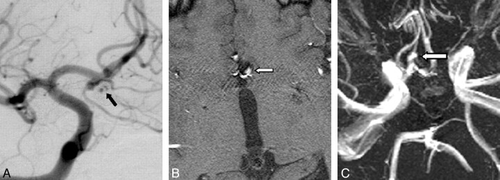
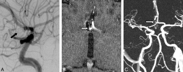
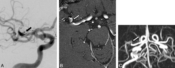
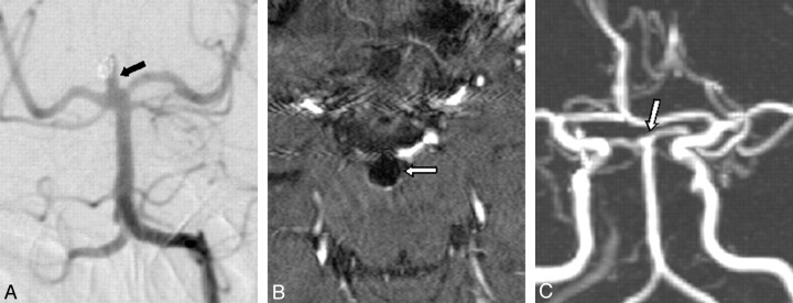
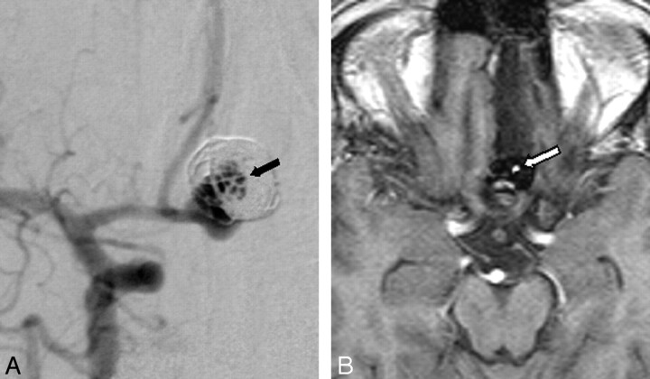
Background and purpose: 3D time-of-flight MR angiography (3D TOF MRA) may be used as noninvasive alternative to digital subtraction angiography (DSA) for the follow-up of patients with intracranial aneurysms treated with Guglielmi detachable coils (GDCs). We aimed to determine the influence of aneurysm size and location on diagnostic accuracy of 3D TOF MRA for follow-up of intracranial aneurysms treated with GDCs.
Materials and methods: Two hundred and one 3D TOF MRAs in 127 consecutive patients with 136 aneurysms were compared with DSA as standard of reference. Sensitivity and specificity of 3D TOF MRA for detection of residual or reperfusion of the aneurysms was calculated with regard to aneurysm size and location.
Results: Overall sensitivity and specificity of MRA was 88.5% and 92.9%, respectively. Sensitivity was lower for aneurysms <or=5 mm (72.2%) and <or=3 mm (63.6%). In addition to the small aneurysm size, interpretation of MR angiograms was compromised by susceptibility artifacts at the air-bone interface, arterial overlap, and pulsation-induced artifacts. The small number of disagreements between MRA and DSA hampered reliable interpretation of the possible influence of aneurysm location on MRA accuracy.
Conclusion: The sensitivity of 3D TOF MRA for detection of reperfusion or residual perfusion of coiled intracranial aneurysms varies considerably depending on the size of the aneurysms. No conclusions can be drawn regarding a possible influence of aneurysm location on diagnostic accuracy of 3D TOF MRA. These findings may influence the decision about whether to replace DSA by 3D TOF MRA for the follow-up of patients with intracranial aneurysms treated with GDCs.
Figures
Similar articles
-
Three-dimensional time-of-flight MR angiography for evaluation of intracranial aneurysms after endosaccular packing with Guglielmi detachable coils: comparison with 3D digital subtraction angiography.Eur Radiol. 2004 Jul;14(7):1162-8. doi: 10.1007/s00330-004-2277-5. Epub 2004 Apr 21. Eur Radiol. 2004. PMID: 15103499
-
Time-of-flight MR angiography targeted to coiled intracranial aneurysms is more sensitive to residual flow than is digital subtraction angiography.AJNR Am J Neuroradiol. 2004 Aug;25(7):1154-7. AJNR Am J Neuroradiol. 2004. PMID: 15313700 Free PMC article.
-
Follow-up of intracranial aneurysms selectively treated with coils: Prospective evaluation of contrast-enhanced MR angiography.AJNR Am J Neuroradiol. 2006 Apr;27(4):744-9. AJNR Am J Neuroradiol. 2006. PMID: 16611757 Free PMC article. Clinical Trial.
-
MRA versus DSA for follow-up of coiled intracranial aneurysms: a meta-analysis.AJNR Am J Neuroradiol. 2014 Sep;35(9):1655-61. doi: 10.3174/ajnr.A3700. Epub 2013 Sep 5. AJNR Am J Neuroradiol. 2014. PMID: 24008171 Free PMC article.
-
Diagnosing flow residuals in coiled cerebral aneurysms by MR angiography: meta-analysis.J Neurol. 2014 Apr;261(4):655-62. doi: 10.1007/s00415-013-7053-5. Epub 2013 Jul 28. J Neurol. 2014. PMID: 23893001 Free PMC article. Review.
Cited by
-
Magnetic Resonance Angiography in the Diagnosis of Cerebral Arteriovenous Malformation and Dural Arteriovenous Fistulas: Comparison of Time-Resolved Magnetic Resonance Angiography and Three Dimensional Time-of-Flight Magnetic Resonance Angiography.Iran J Radiol. 2016 Mar 28;13(2):e19814. doi: 10.5812/iranjradiol.19814. eCollection 2016 Apr. Iran J Radiol. 2016. PMID: 27679690 Free PMC article.
-
Features of "false positive" unruptured intracranial aneurysms on screening magnetic resonance angiography.PLoS One. 2020 Sep 3;15(9):e0238597. doi: 10.1371/journal.pone.0238597. eCollection 2020. PLoS One. 2020. PMID: 32881975 Free PMC article.
-
Three-dimensional time-of-flight MR angiography at 3 T compared to digital subtraction angiography in the follow-up of ruptured and coiled intracranial aneurysms: a prospective study.Neuroradiology. 2008 May;50(5):383-9. doi: 10.1007/s00234-007-0355-5. Neuroradiology. 2008. PMID: 18196229 Clinical Trial.
-
Highly accelerated intracranial time-of-flight magnetic resonance angiography using wave-encoding.Magn Reson Med. 2023 Aug;90(2):432-443. doi: 10.1002/mrm.29647. Epub 2023 Apr 3. Magn Reson Med. 2023. PMID: 37010811 Free PMC article.
-
Follow-up of coiled cerebral aneurysms at 3T: comparison of 3D time-of-flight MR angiography and contrast-enhanced MR angiography.AJNR Am J Neuroradiol. 2008 Sep;29(8):1530-6. doi: 10.3174/ajnr.A1166. Epub 2008 Jun 12. AJNR Am J Neuroradiol. 2008. PMID: 18556359 Free PMC article. Clinical Trial.
References
-
- Brunereau L, Cottier JP, Sonier CB, et al. Prospective evaluation of time-of-flight MR angiography in the follow-up of intracranial saccular aneurysms treated with Guglielmi detachable coils. J Comput Assist Tomogr 1999;23:216–23 - PubMed
-
- Boulin A, Pierot L. Follow-up of intracranial aneurysms treated with detachable coils: comparison of gadolinium-enhanced 3D time-of-flight MR angiography and digital subtraction angiography. Radiology 2001;219:108–13 - PubMed
-
- Weber W, Yousry TA, Felber SR, et al. Noninvasive follow-up of GDC-treated saccular aneurysms by MR angiography. Eur Radiol 2001;11:1792–97 - PubMed
Publication types
MeSH terms
Substances
LinkOut - more resources
Full Text Sources
Medical