

Correlation of diffusion tensor and dynamic perfusion MR imaging metrics in normal-appearing corpus callosum: support for primary hypoperfusion in multiple sclerosis
- PMID: 17416836
- PMCID: PMC7977353
Correlation of diffusion tensor and dynamic perfusion MR imaging metrics in normal-appearing corpus callosum: support for primary hypoperfusion in multiple sclerosis
Abstract
Background and purpose: Hypoperfusion of the normal-appearing white matter in multiple sclerosis (MS) may be related to ischemia or secondary to hypometabolism from wallerian degeneration (WD). This study evaluated whether correlating perfusion and diffusion tensor imaging (DTI) metrics in normal-appearing corpus callosum could provide support for an ischemic mechanism for hypoperfusion.
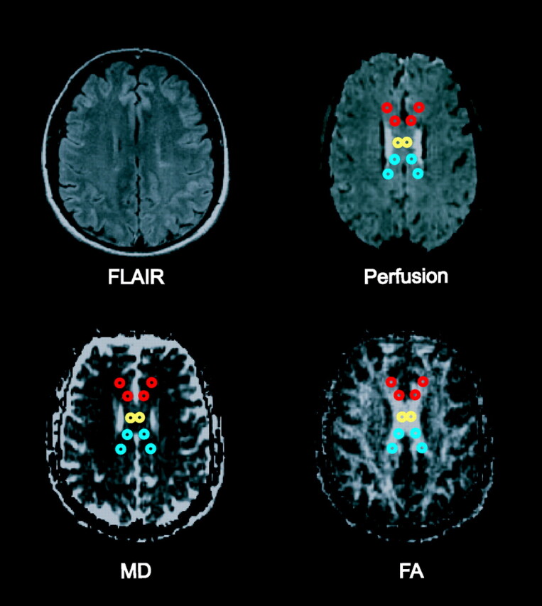
Materials and methods: Fourteen patients with relapsing-remitting MS (RRMS) and 17 control subjects underwent perfusion MR imaging and DTI. Absolute measures of cerebral blood volume (CBV), cerebral blood flow (CBF), and mean transit time (MTT) were calculated. Mean diffusivity (MD) and fractional anisotropy (FA) maps were computed from DTI data. After visual coregistration of perfusion and DTI images, regions of interest were placed in the genu, central body, and splenium of normal-appearing corpus callosum. Pearson product-moment correlation coefficients were calculated using mean DTI and perfusion measures in each region.
Results: In the RRMS group, CBF and CBV were significantly correlated with MD in the splenium (r = 0.83 and r = 0.63, respectively; both P < .001) and in the central body (r = 0.86 and r = 0.65, respectively; both P < .001), but not in the genu (r = 0.23 and 0.25, respectively; both P is nonsignificant). No significant correlations were found between MTT and DTI measures or between FA and any perfusion measure in the RRMS group. No significant correlations between diffusion and perfusion metrics were found in control subjects.
Conclusion: In the normal-appearing corpus callosum of patients with RRMS, decreasing perfusion is correlated with decreasing MD. These findings are more consistent with what would be expected in primary ischemia than in secondary hypoperfusion from WD.
Figures
References
-
- Trotter JL, Wegescheide CL, Garvey WF, et al. Studies of myelin proteins in multiple sclerosis brain tissue. Neurochem Res 1984;9:147–52 - PubMed
-
- Trapp BD, Peterson J, Ransohoff RM, et al. Axonal transection in the lesions of multiple sclerosis. N Engl J Med 1998;338:278–85 - PubMed
-
- Traugott U, Reinherz EL, Raine CS. Multiple sclerosis. Distribution of T cells, T cell subsets and Ia-positive macrophages in lesions of different ages. J Neuroimmunol 1983;4:201–21 - PubMed
-
- Adams CW. Pathology of multiple sclerosis: progression of the lesion. Br Med Bull 1977;33:15–20 - PubMed
-
- Narayanan S, Fu L, Pioro E, et al. Imaging of axonal damage in multiple sclerosis: spatial distribution of magnetic resonance imaging lesions. Ann Neurol 1997;41:385–91 - PubMed
MeSH terms
Substances
LinkOut - more resources
Full Text Sources
Medical