

Phospholipase A2 in chamber angle of normal eyes and patients with primary open angle glaucoma and exfoliation glaucoma
- PMID: 17417602
- PMCID: PMC2642936
Phospholipase A2 in chamber angle of normal eyes and patients with primary open angle glaucoma and exfoliation glaucoma
Abstract
Purpose: Phospholipase A2 (PLA2) is a growing family of lipolytic enzymes that play a key role in various biological processes including general lipid metabolism, membrane homeostasis, and in diseases such as atherosclerosis, arthritis, and acute pancreatitis. Oxidative stress as well as inflammation may be associated with glaucoma pathogenesis. Therefore, our aim was to examine the expression of group IIA secretory PLA2 (sPLA2-IIA), group V secretory PLA2 (sPLA2-V), calcium-independent PLA2 (iPLA2), and cytosolic PLA2 (cPLA2) type in the trabecular meshwork (TM) and the canal of Schlemm in normal eyes and in juxtacanalicular tissue samples from patients with primary open angle glaucoma (POAG) or exfoliation glaucoma (ExG).
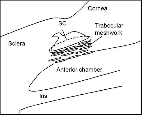
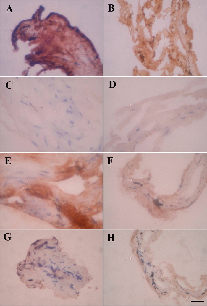
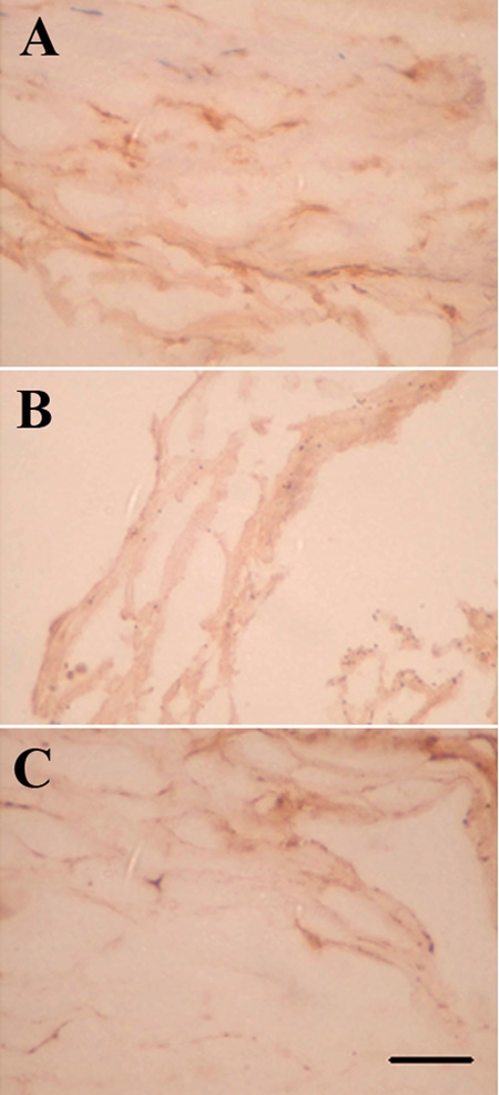
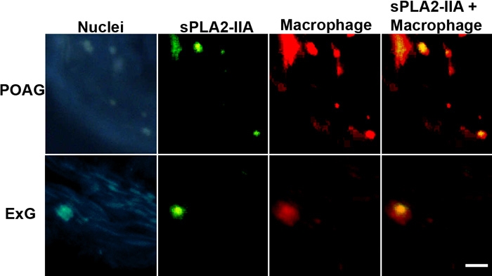
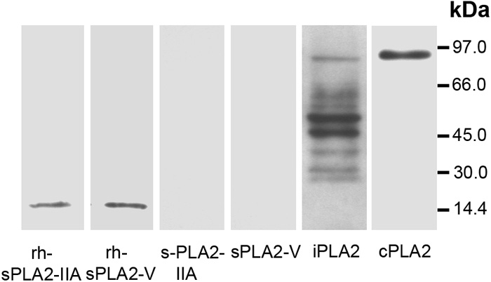
Methods: TM tissues were isolated from healthy donor eyes for corneal transplantation. Specimens of inner wall of the Schlemm's canal and the juxtacanalicular tissue were collected during deep sclerectomy from the eyes of patients who had POAG or ExG. Antibodies against PLA2s (sPLA2-IIA, sPLA2-V, iPLA2, and cPLA2) and a standard immunohistochemical procedure were used for the analysis. Quantification of immunoreactions was provided using a Photoshop-based image analysis. Double-staining immunofluorescence of macrophages and sPLA2-IIA was performed by using confocal microscopy.
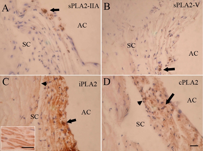
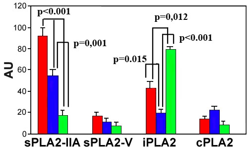
Results: sPLA2-IIA was not present in normal TM. In contrast, sPLA2-IIA levels were significantly higher in glaucoma patients than in controls. Furthermore, sPLA2-IIA expression was much higher in POAG when compared to ExG. iPLA2 was found to predominate in normal human TM, and it demonstrated strong labeling in the uveal and corneoscleral meshwork. The staining of juxtacanalicular meshwork was only moderate in density. In contrast, expression of the enzyme was significantly decreased in glaucoma patients, especially in ExG, when compared to normal controls or to POAG. In addition, strong regional differences were detected in sPLA2-IIA and iPLA2 levels in POAG, whereas immunostaining of these enzymes was much lower and rather uniform throughout ExG sample. In POAG, sPLA2-IIA staining was restricted to certain parts of the trabecular samples where sPLA2-IIA positive macrophages were also present. Immunostaining of sPLA2-V or cPLA2 was low, and no significant changes were found in levels of these enzymes between normal and glaucomatous samples.
Conclusions: sPLA2-IIA, an oxidative stress marker in atherosclerosis, is overexpressed especially in POAG. This result supports the hypothesis that oxidative stress may play a significant role in the pathogenesis of POAG. In ExG, a dramatic decrease in the expression level of iPLA2, a housekeeping enzyme in phospholipid remodeling, may indicate imbalance in phospholipid turnover and also inhibition of normal physiological functions in the TM. These findings may contribute to understanding the pathogenesis of POAG and ExG and may be important for the development of novel therapeutic strategies to different glaucomas.
Figures
References
-
- Weinreb RN, Khaw PT. Primary open-angle glaucoma. Lancet. 2004;363:1711–20. - PubMed
-
- Coleman AL. Glaucoma. Lancet. 1999;354:1803–10. - PubMed
-
- Forsius H. Prevalence of pseudoexfoliation of the lens in Finns, Lapps, Icelanders, Eskimos, and Russians. Trans Ophthalmol Soc U K. 1979;99:296–8. - PubMed
-
- Krause U, Helve J, Forsius H. Pseudoexfoliation of the lens capsule and liberation of iris pigment. Acta Ophthalmol (Copenh) 1973;51:39–46. - PubMed
Publication types
MeSH terms
Substances
LinkOut - more resources
Full Text Sources