

Atomic force microscopy measurements of lens elasticity in monkey eyes
- PMID: 17417612
- PMCID: PMC2649306
Atomic force microscopy measurements of lens elasticity in monkey eyes
Abstract
Purpose: To demonstrate the feasibility of measuring the elasticity of intact crystalline lenses using atomic force microscopy (AFM).
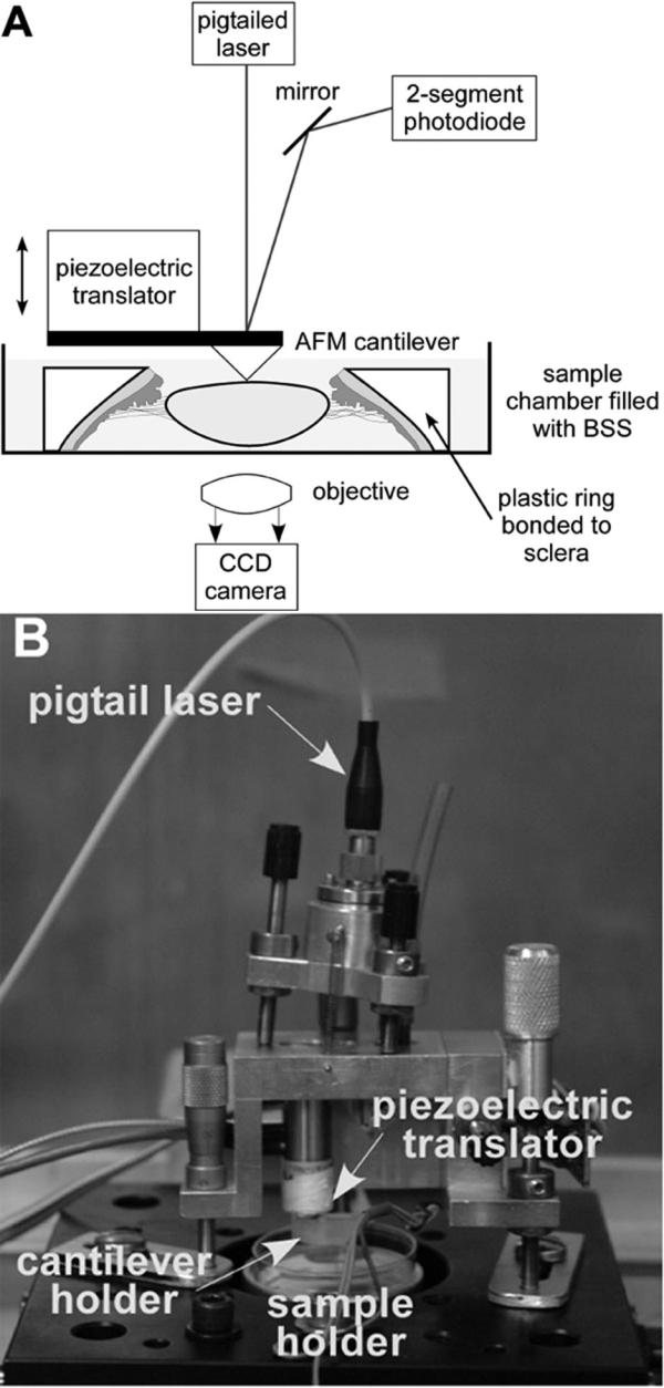
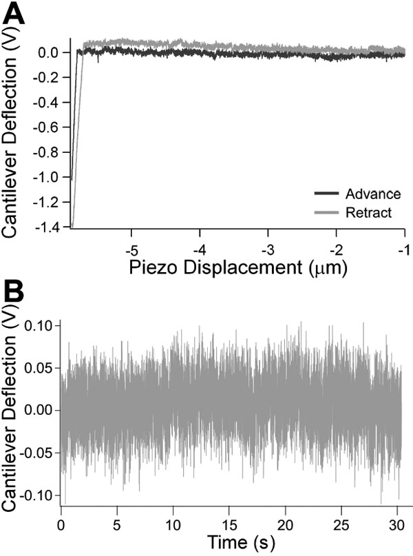
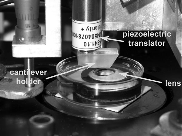
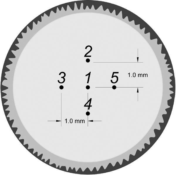
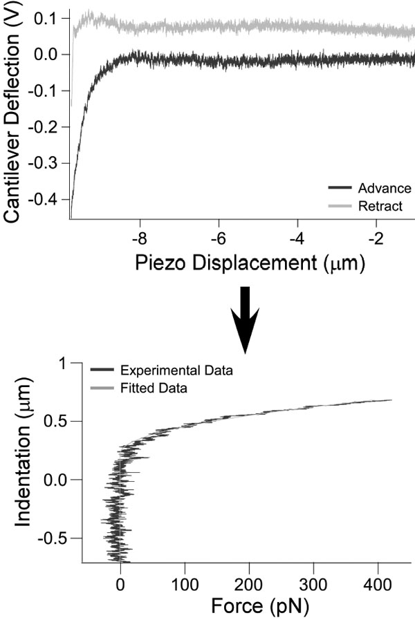
Methods: AFM elasticity measurements were performed on intact lenses from 18 fresh cynomolgus monkey cadaver eyes (4-10 years old, <1 day postmortem) that had been left attached to their zonule-ciliary body-sclera framework. The eyes were prepared by bonding a plastic ring on the sclera after removal of the conjunctival, adipose, and muscle tissues. The posterior pole was sectioned, with the excess vitreous removed, and the eye's anterior section was placed on a Teflon slide to protect the posterior pole of the lens. The cornea and iris were then sectioned. The lens-zonule-ciliary body-sclera section was then placed in a Petri dish filled with balanced salt solution in an AFM system designed for force measurements. Next, the central pole of the anterior surface of the intact lens was probed with the AFM cantilever tip. The recorded AFM cantilever deflection-indentation curves were used to derive force-indentation curves for the lens after factoring out the deflection of the cantilever on a hard surface. Young's modulus of the lens was calculated from the force-indentation relation using the Hertz model.
Results: Young's modulus was 1,720+/-880 Pa (range: 409-3,210 Pa) in the 18 cynomolgus monkey lenses.
Conclusions: AFM can be used to provide measurements of the elasticity of the whole lens including the capsule. Values obtained using AFM on cynomolgus monkey lenses are similar to published values obtained using dynamic mechanical analysis on young human lenses.
Figures
Comment in
-
Lens hardness not related to the age-related decline of accommodative amplitude.Mol Vis. 2007 Jun 27;13:1010-1. Mol Vis. 2007. PMID: 17653044 Free PMC article. Review. No abstract available.
References
-
- Fincham EF. The Mechanism of Accommodation. British Journal of Ophthalmology. 1937;Monograph Supplement VIII
-
- Wyatt HJ. Some aspects of the mechanics of accommodation. Vision Res. 1988;28:75–86. - PubMed
-
- Glasser A, Kaufman PL. Accommodation and Presbyopia. In: Kaufman PL, Alm A, editors. Adler's Physiology of the Eye. 10th ed. St Louis: Mosby; 2003. p. 195-233.
-
- Strenk SA, Strenk LM, Koretz JF. The mechanism of presbyopia. Prog Retin Eye Res. 2005;24:379–93. - PubMed
Publication types
MeSH terms
Grants and funding
LinkOut - more resources
Full Text Sources
Other Literature Sources
Miscellaneous