. 2007 Jul 1;463(1):60-7.
doi: 10.1016/j.abb.2007.02.030. Epub 2007 Mar 16.

Assembly of recently translated full-length and C-terminal truncated human gamma-globin chains with a pool of alpha-globin chains to form Hb F in a cell-free system

Affiliations

Assembly of recently translated full-length and C-terminal truncated human gamma-globin chains with a pool of alpha-globin chains to form Hb F in a cell-free system

Kazuhiko Adachi et al. Arch Biochem Biophys. .

Abstract

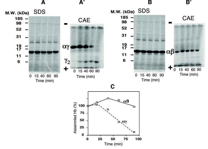
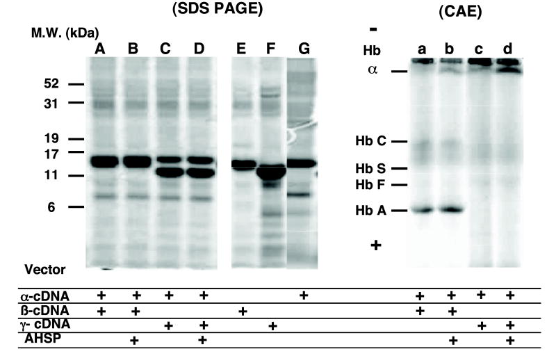
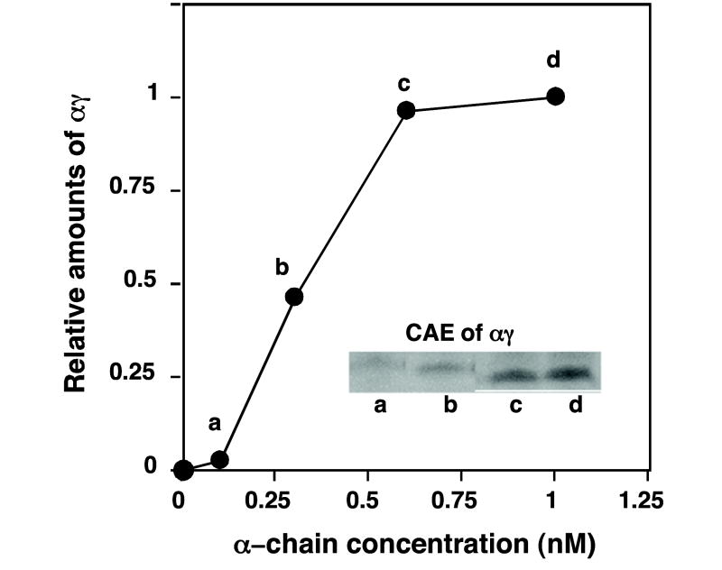
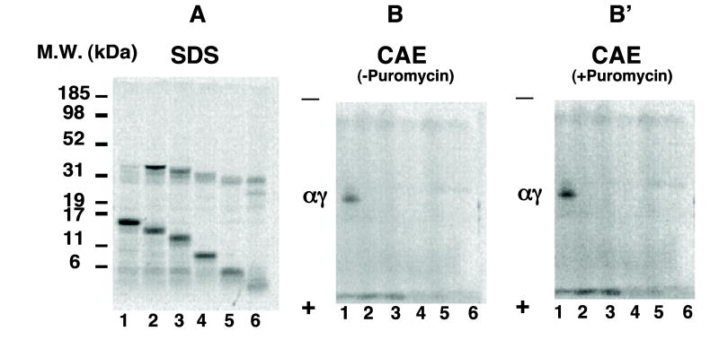
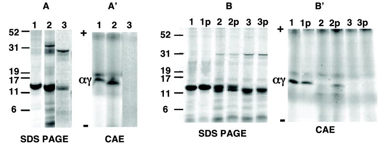
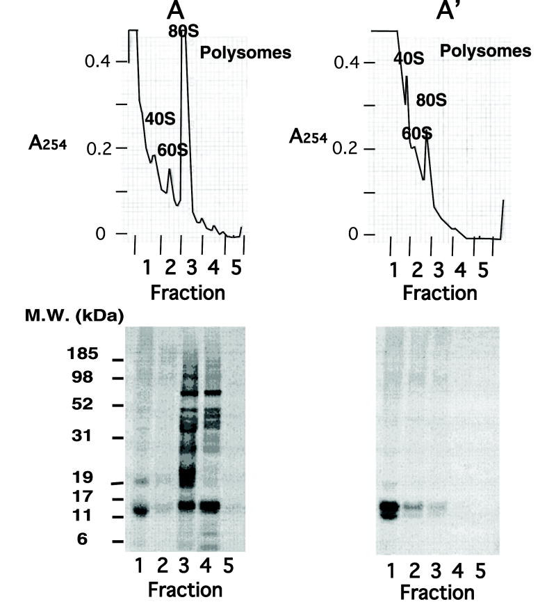
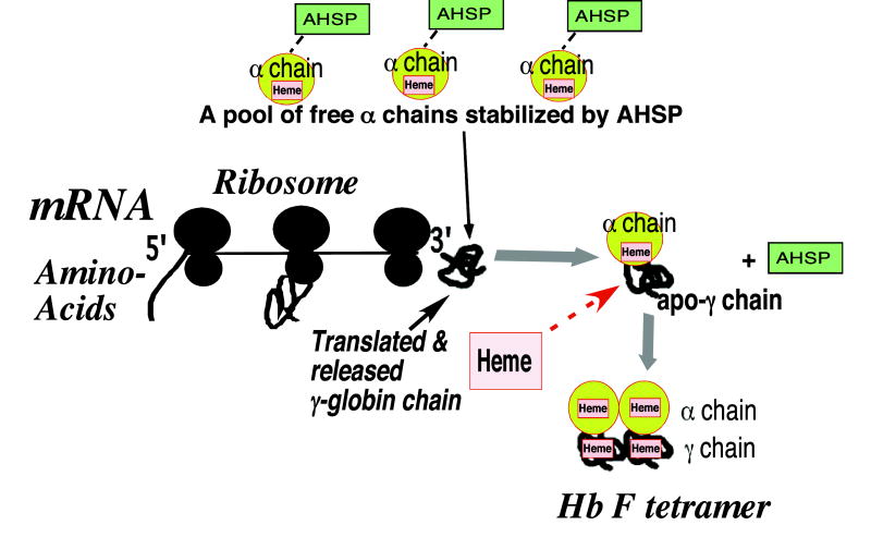
Assembly of alpha-globin with translated, full-length and C-terminal truncated human gamma-globin to form Hb F was assessed in a cell-free transcription/translation system. Polysome profiles showed two amino acid C-terminal-truncated gamma-chains retained on polysomes can assemble with unlabeled holo alpha-chains only after puromycin-induced chain release. Two amino acid C-terminal truncated gamma-chains encoded from vectors containing a stop codon at the translation termination site were released from polysomes and assembled with alpha-chains in the absence of puromycin addition, while removal of 11 or more amino acids from the gamma-chain carboxy-terminus inhibited assembly with alpha-chains. These results suggest that amino acids in the HC- and H-helix gamma-chain regions including amino acids 135-144 at the C-terminus in the translated gamma-chains play a key role in assembly with alpha-chains, and that assembly occurs soon after exit of translated gamma-chains from the ribosome tunnel and release from polysomes thereby preventing stable gamma(2) homo-dimer formation.


Figures

Fig. 1
Fig. 1
Assembly of radiolabeled γ- or β-globin chains following addition of unlabeled α-globin chains at different times during reactions in a cell-free coupled transcription/translation system. Transcription/translation of γ-globin cDNA and β-globin cDNA vectors was performed following addition of unlabeled α-globin chains at different times during the 90 min reactions. Time of addition of unlabeled α chains (7.5 nM) is shown on the x-axis, and radiolabeled globin chains synthesized after 90 min are shown after SDS–PAGE ((A) and (B) are results for γ- and β-globin chains, respectively). Assembled homo-dimers and tetramers are shown following cellulose electrophoresis (CAE) (in A′ and B′ for αγ and αβ formation, respectively, or formation of their homo-dimers). Relative amounts of hetero-dimer formation for αβ and αγ as a function of time of addition of unlabeled α-globin chains during the 90 min. reaction are shown in (C).
Fig. 2
Fig. 2
Globin-chain synthesis and tetramer formation from α and non-α globin chains co-expressed in the same cell-free coupled transcription/translation reaction. Radiolabeled globin chains synthesized after a 60-min reaction containing only one expression vector (lanes E, F, and G) or equal amounts of α and non-α chain cDNA expression vectors (lanes A–D) in the presence (lanes B and D) or absence (lanes A, C, E, F and G) of AHSP (~10 nM) is shown after SDS–PAGE and autoradiography (left panel). Assembled radiolabeled hemoglobin tetramers formed from reactions corresponding to the left panel (lanes a–d) are shown after CAE and autoradiography after addition of unlabeled Hb A or Hb F (~1.6 nM) prior to electrophoresis. Positions for migration of Hbs A, F, S and C as well as free α globin are indicated.
Fig. 3
Fig. 3
Effects of varying unlabeled α-globin chain amounts added at zero time on formation of radiolabeled hetero-dimers. Radiolabeled αγ formation is shown after 30 min reactions as a function of unlabeled α chain amount added at 0 time. Reactions were subjected to CAE followed by autoradiography. Relative radiolabeled αγ band intensities were quantitated following autoradiography (inset) using a phosphorimaging analysis system (STORM 84D), and results expressed as relative amounts where 1.0 on the y-axis represents value obtained with 1nM unlabeled α chains added at zero time to reactions.
Fig. 4
Fig. 4
Assembly of truncated γ chains templated from linearized cDNA expression vectors lacking a stop codon with α chains in a cell-free coupled transcription/translation system. A variety of restrictive enzymes (HindIII, MscI, EcoRI, PvuII, and NcoI) was used to generate linearized expression vector templates terminated at varying lengths from 24% to 100% (146 amino acids) of wild-type template. Transcription/translation of truncated, linearized γ-globin cDNA expression vectors was performed for 30 min following addition of unlabeled α-globin chains (7.5 nM) at zero time. Synthesis of radiolabeled globin chains (SDS–PAGE, left panel A) and assembled hetero-dimers from reactions containing (CAE, right panel B′) or lacking (CAE, middle panel B) puromycin (5 mM) are shown. Lanes 1, 2, 3, 4, 5 and 6 represent results for γ-chain templates containing 146 (wild type), 135, 119, 86, 58 or 35 amino acids.
Fig. 5
Fig. 5
Assembly of two amino acid-truncated γ-globin chains from reactions containing or lacking puromycin. KpnI or MscI were used to generate linearized expression vector templates terminated after 144 and 135 amino acids in the γ chain. These templates lack a stop codon and were compared to γ-chain expression vectors containing PCR-induced stop codons after 135 or 144 amino acids. Transcription/translation reactions of these expression vector templates containing (A and A′) or lacking (B and B′) a stop codon were performed for 30 min following addition of unlabeled α-globin chains (7.5 nM) at zero time. Results were compared to those of wild type γ-globin cDNA. Synthesis of radiolabeled globin chains are shown after SDS–PAGE from templates containing (A) or lacking (B) a stop codon. Corresponding assembled hetero-dimers are shown after CAE from templates containing (A′) or lacking (B′) a stop codon. Lanes 2 and 3 represent results from two or eleven amino-acid truncated versus wild γ-cDNA expression vector (lane 1), respectively. Lane 2p and 3p in (B) and (B′) represent results for two or eleven amino acid truncated and wild γ-cDNA reactions (lane 1p), respectively, incubated with puromycin (5 mM).
Fig. 6
Fig. 6
Polysome profiles of reactions containing KpnI-linearized two amino acid truncated γ-chain cDNA vector incubated with α chains in the presence or absence of puromycin. Transcription/translation reactions containing truncated templates were incubated at 30 °C in the presence of unlabeled α chains (7.5 nM) added at zero time. After 30 min, the reaction was incubated for an additional 10 min in the presence (A′) or absence (A) of puromycin (5 mM) at room temperature. Reactions were fractionated on sucrose gradients and fractions were collected. Absorbance at 254 nm is shown in the top panels (A and A′), while results of SDS–PAGE followed by autoradiography are shown below. Polysomal fractions are in tubes 3–5.
Fig. 7
Fig. 7
Diagram of proposed assembly of recently translated γ-globin chains with AHSP- stabilized α chains to form Hb F in vivo. The nascently growing apo-γ globin polypeptide chains are shown during translation forming partially folded structures prior to complete translation. A pool of α chains stabilized by binding to AHSP interacts with recently translated, full-length apo-γ-globin chains dynamically after their exit from the ribosome tunnel and release generating α-apo-γ hetero-dimers and free AHSP, which results in prevention of γ2 homo-dimer formation. Assembled α-apo-γ hetero-dimers promote correct heme insertion into the heme pockets of the γ chains. In addition, α chains may act as chaperones and promote formation of stable αγ hetero-dimers by facilitating proper conformational folding of γ-globin chains during assembly.

Similar articles

References

    1. Bunn FH. Blood. 1987;69:1–6. - PubMed
    1. Shaeffer JR, Kingston RE, McDonald MJ, Bunn HF. Nature. 1978;276:631–632. - PubMed
    1. Adachi K, Zhao Y, Yamaguchi T, Surrey S. J Biol Chem. 2000;275:12424–12429. - PubMed
    1. Adachi K, Zhao Y, Surrey S. J Biol Chem. 2002;277:13415–13420. - PubMed
    1. Stalling M, Abraham A, Abraham EC. Blood. 1983;62:75a. - PubMed

Publication types

LinkOut - more resources