

Rebuilding the coronary vasculature: hedgehog as a new candidate for pharmacologic revascularization
- PMID: 17418368
- PMCID: PMC2267919
- DOI: 10.1016/j.tcm.2007.01.002
Rebuilding the coronary vasculature: hedgehog as a new candidate for pharmacologic revascularization
Abstract
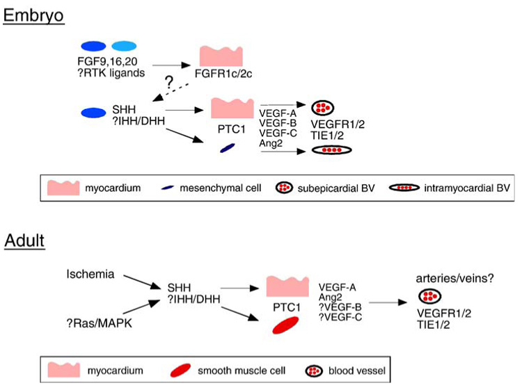
Myocardial infarction and ischemic heart disease are among the most common causes of morbidity and mortality in the industrial world. Surgical and percutaneous intravascular approaches are commonly used to treat these diseases. Regrettably, a significant number of patients are either ineligible or demonstrate suboptimal responses to these therapies. In an attempt to provide such patients improved therapeutic options, much effort has been spent developing noninvasive approaches to restore coronary vascular perfusion. One such strategy, termed therapeutic revascularization or angiogenesis, involves administration of proangiogenic factors, which improve coronary perfusion by promoting growth of the coronary vasculature. Thus far, two potential proangiogenic factors have been intensively examined, fibroblast growth factor and vascular endothelial growth factor. Unfortunately, despite their apparent efficacy in animal models, neither factor has performed adequately in the clinic to date. Within the past year a new factor, hedgehog, has been shown to effectively promote the growth of the coronary vasculature and thus has been proposed as a novel candidate for therapeutic revascularization. In this review, we discuss the discovery of the hedgehog pathway as an essential regulator of the development of the coronary vasculature, as an inducer of adult coronary vascular growth, and as a therapeutic in the treatment of ischemic heart disease.
Figures


References
-
- Asai J, Takenaka H, Kusano KF, et al. Topical sonic hedgehog gene therapy accelerates wound healing in diabetes by enhancing endothelial progenitor cell-mediated microvascular remodeling. Circulation. 2006;113:2413–2424. - PubMed
-
- Auguste P, Javerzat S, Bikfalvi A. Regulation of vascular development by fibroblast growth factors. Cell Tissue Res. 2003;314:157–166. - PubMed
-
- Caines AE, Massad MG, Kpodonu J, et al. Outcomes of coronary artery bypass grafting versus percutaneous coronary intervention and medical therapy for multi-vessel disease with and without left ventricular dysfunction. Cardiology. 2004;101:21–28. - PubMed
-
- Chen TH, Chang TC, Kang JO, et al. Epicardial induction of fetal cardiomyocyte proliferation via a retinoic acid-inducible trophic factor. Dev Biol. 2002;250:198–207. - PubMed
-
- Davies R, Moore A, Schedl A, et al. Multiple roles for the Wilms’ tumor suppressor WT1. Cancer Res. 1999;59:1747s–1750s. [discussion 1751s] - PubMed
Publication types
MeSH terms
Substances
Grants and funding
LinkOut - more resources
Full Text Sources
Other Literature Sources
Miscellaneous
