. 2007 May;44(12):3092-9.
doi: 10.1016/j.molimm.2007.02.009. Epub 2007 Apr 6.

Beta-arrestins 1 and 2 differentially regulate LPS-induced signaling and pro-inflammatory gene expression

Affiliations

Beta-arrestins 1 and 2 differentially regulate LPS-induced signaling and pro-inflammatory gene expression

Hongkuan Fan et al. Mol Immunol. 2007 May.

Abstract

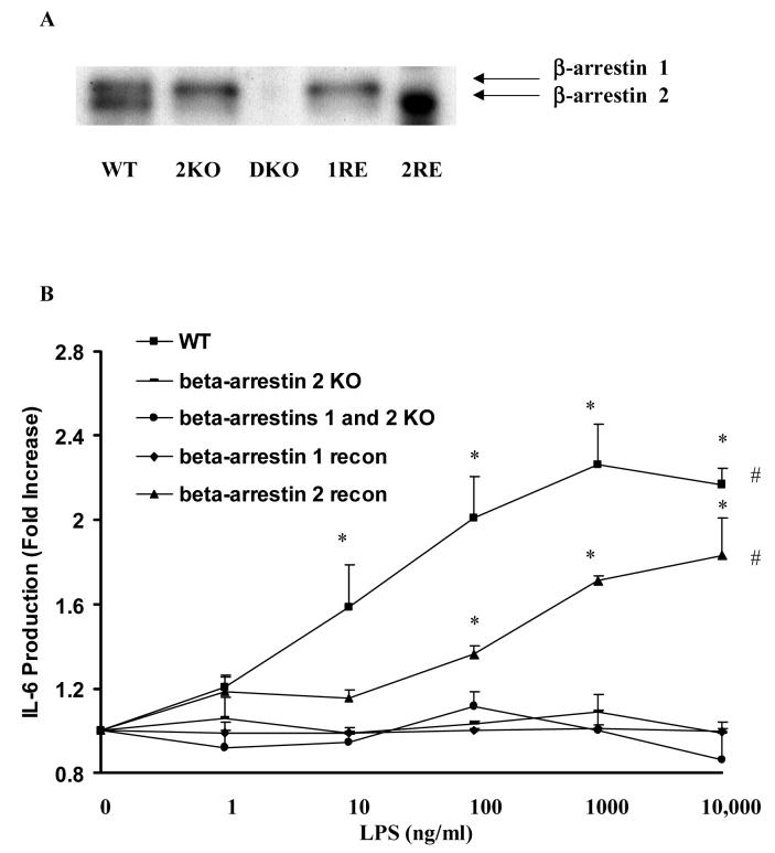
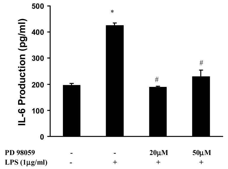
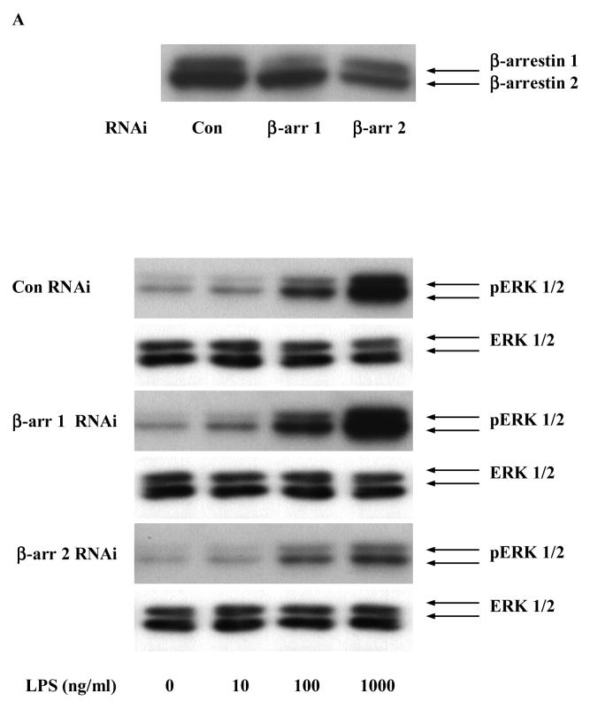
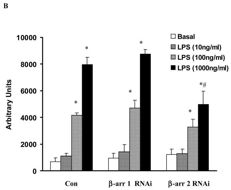
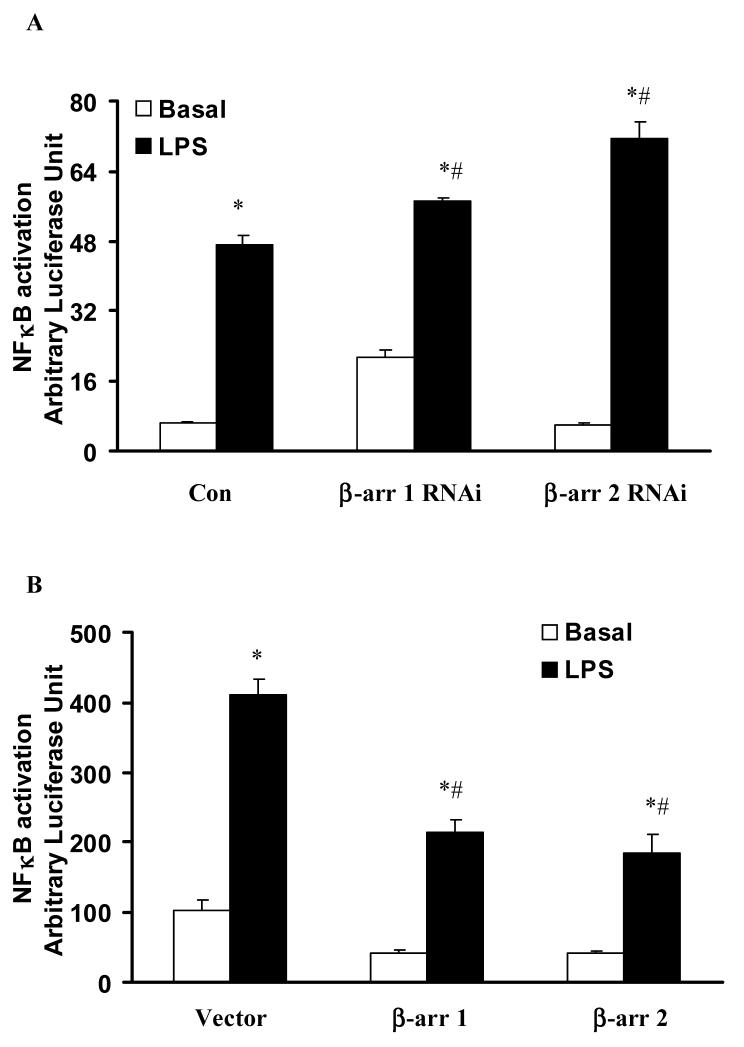
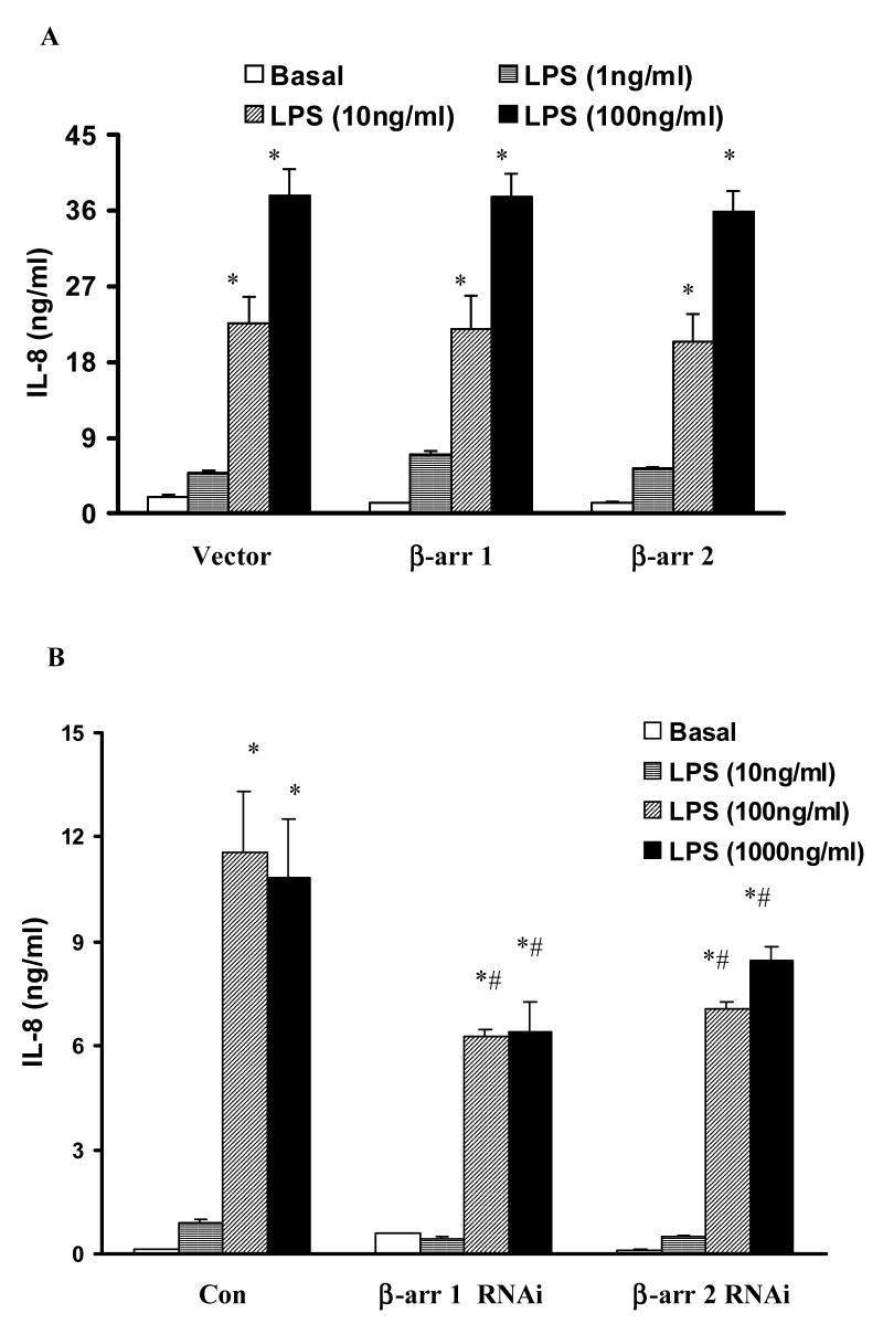
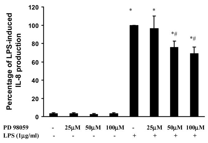
Toll like receptors, the critical receptor family in innate immunity, have been shown to signal via both ERK 1/2 and transcription factor NFkappaB. beta-Arrestins 1 and 2 have recently been implicated in modulation of NFkappaB signaling and ERK 1/2 activation. Using a number of approaches: mouse embryonic fibroblasts (MEF) from wild-type (WT), beta-arrestins knockouts (KO), beta-arrestins 1 and 2 double KO, and MEFs with reconstituted WT beta-arrestins in the double KO cells, RNA interference (siRNA) specific knockdown of beta-arrestins, and overexpression of WT beta-arrestins, it was demonstrated that beta-arrestin 2 positively regulates LPS-induced ERK 1/2 activation and both beta-arrestins 1 and 2 negatively regulate LPS-induced NFkappaB activation. Also beta-arrestin 2 positively regulate LPS-induced IL-6 production and both beta-arrestins 1 and 2 positively regulate LPS-induced IL-8 production. The specific ERK1/2 inhibitor PD98059 significantly decreased LPS-induced IL-6 and IL-8 production suggesting that IL-6 and IL-8 production is, in part, mediated by ERK 1/2 activation. Over expression of wild type beta-arrestins 1 and 2 had no effect on LPS-induced ERK1/2 activation and LPS-induced IL-8 production suggesting that endogenous beta-arrestins 1 and 2 are sufficient to mediate maximum ERK 1/2 activity and IL-8 production. beta-Arrestins thus not only negatively regulate LPS-induced NFkappaB activation but also positively regulate ERK 1/2 activation and specific pro-inflammatory gene expression. Understanding the role of beta-arrestins in regulation of TLR signaling pathways may provide novel insights into control mechanisms for inflammatory gene expression.


Figures

Figure 1
Figure 1. Effect of altered β-arrestins 1/2 expression on LPS-induced IL-6 production in mouse embryonic fibroblasts
MEFs from WT mice, β-arrestin 2 knockout mice, β-arrestin 1/2 double knockout mice and re-constitution of wild-type β-arrestin 1 or 2 in the double knockout MEF cells were stimulated with LPS (0-10μg/ml). LPS-induced IL-6 production was studied (A). β-arrestin 1 and 2 expression in these MEF cell lines were examined by Western blot analysis (B). Data represent means ± SE from three independent experiments. *, p<0.05 compared to naïve cells; #, p<0.05 compared to β-arrestin 2 KO, β-arrestin 1/2 DKO, and β-arrestin 1 re-constituted MEF cells.
Figure 2
Figure 2. Effect of PD98059 on LPS-induced IL-6 production in MEFs
MEFs from WT mice were pretreated with PD98059 (20μM-50μM) a specific ERK1/2 inhibitor followed by stimulation with LPS (1μg/ml) for 18 hours. Data represent means ± SE from three independent experiments. *, p<0.05 compared to basal cells; #, p<0.05 compared to LPS stimulated groups.
Figure 3
Figure 3. Effect of β-arrestins 1/2 RNAi on LPS-induced ERK 1/2 activation in HEK-TLR4 cells
HEK 293 cells stably expressing TLR4, CD14 and MD-2 were transfected with RNAi for control, β-arrestin 1 and β-arrestin 2 followed by stimulation with LPS (0-1μg/ml). LPS-induced ERK1/2 activation was studied (A). Statistical analysis was performed on the densitometry results (B). Data represent means ± SE from three independent experiments. *, p<0.05 compared to basal cells; #, p<0.05 compared to LPS stimulated groups.
Figure 4
Figure 4. Effect of altered β-arrestins expression on LPS-induced NFκB activation in HEK-TLR4 cells
HEK 293 cells stably expressing TLR4, CD14 and MD-2 were transfected with RNAi for control, β-arrestin 1 or β-arrestin 2 (A) or empty vector or wild-type β-arrestin 1 or β-arrestin 2 (B) followed by stimulation with LPS (1μg/ml). LPS-induced NFκB activation was studied. Data represent means ± SE from three independent experiments. *, p<0.05 compared to basal cells; #, p<0.05 compared to LPS stimulated groups.
Figure 5
Figure 5. Effect of altered β-arrestins expression on LPS-induced IL-8 production in HEK-TLR4 cells
HEK 293 cells stably expressing TLR4, CD14 and MD-2 were transfected with empty vector or wild-type β-arrestin 1 or β-arrestin 2 (A) or RNAi for control, β-arrestin 1 or β-arrestin 2 (B) followed by stimulation with LPS (0-1μg/ml). LPS-induced IL-8 production was measured. Data represent means ± SE from three independent experiments. *, p<0.05 compared to basal cells; #, p<0.05 compared to LPS stimulated groups.
Figure 6
Figure 6. Effect of PD98059 on LPS-induced IL-8 production in HEK-TLR4 cells
HEK-TLR4 cells were pretreated with PD98059 (25μM-100μM) a specific ERK1/2 inhibitor followed by stimulation with LPS (1μg/ml) for 18 hours. Data represent means ± SE from three independent experiments. *, p<0.05 compared to basal cells; #, p<0.05 compared to LPS stimulated groups.

References

    1. Ahn S, Nelson CD, Garrison TR, Miller WE, Lefkowitz RJ. Desensitization, internalization, and signaling functions of beta-arrestins demonstrated by RNA interference. Pro.c Natl. Acad. Sci. U S A. 2003;100:1740–1744. - PMC - PubMed
    1. Ahn S, Wei H, Garrison TR, Lefkowitz RJ. Reciprocal regulation of angiotensin receptor-activated extracellular signal-regulated kinases by beta-arrestins 1 and 2. J. Biol. Chem. 2004;279:7807–7811. - PubMed
    1. Akashi S, Shimazu R, Ogata H, Nagai Y, Takeda K, Kimoto M, Miyake K. Cutting edge: cell surface expression and lipopolysaccharide signaling via the toll-like receptor 4-MD-2 complex on mouse peritoneal macrophages. J. Immunol. 2000;164:3471–3475. - PubMed
    1. Akira S, Takeda K. Toll-like receptor signalling. Nat. Rev. Immunol. 2004;4:499–511. - PubMed
    1. Akira S, Uematsu S, Takeuchi O. Pathogen recognition and innate immunity. Cell. 2006;124:783–801. - PubMed

Publication types