. 2007;31(2):110-8.
doi: 10.1016/j.cdp.2007.02.005. Epub 2007 Apr 6.

Inhibition of cell proliferation, induction of apoptosis, reactivation of DLC1, and modulation of other gene expression by dietary flavone in breast cancer cell lines

Affiliations

Inhibition of cell proliferation, induction of apoptosis, reactivation of DLC1, and modulation of other gene expression by dietary flavone in breast cancer cell lines

Veronika Ullmannova et al. Cancer Detect Prev. 2007.

Abstract

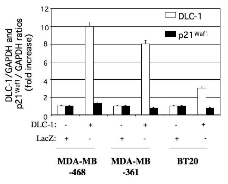
Background: Dietary flavone was previously shown to increase the expression of deleted in liver cancer-1 gene (DLC-1) in HT-29 colon carcinoma cell line [Herzog A, Kindermann B, Doring F, Daniel H, Wenzel U. Pleiotropic molecular effects of the pro-apoptotic dietary constituent flavone in human colon cancer cells identified by protein and mRNA expression profiling. Proteomics 2004;4:2455-64]. DLC-1 that encodes a Rho GTPase-activating protein, functions as a tumor suppressor gene and is frequently inactivated or down-regulated in several common cancers. Restoration of DLC-1 expression suppresses in vitro tumor cells proliferation and tumorigenicity in vivo.

Methods: Here, the effect of flavone was examined in several DLC-1-deficient cell lines derived from different types human cancer using assays for cell proliferation, gene expression and transfer.

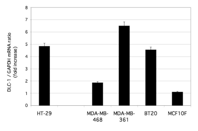
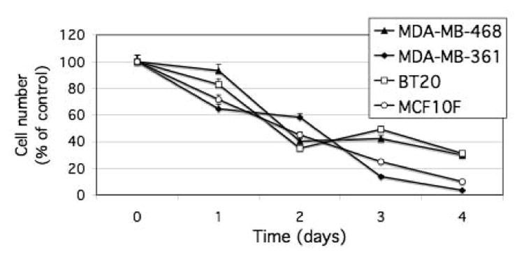
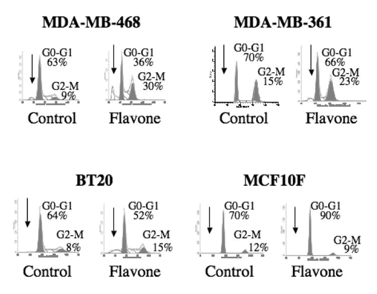
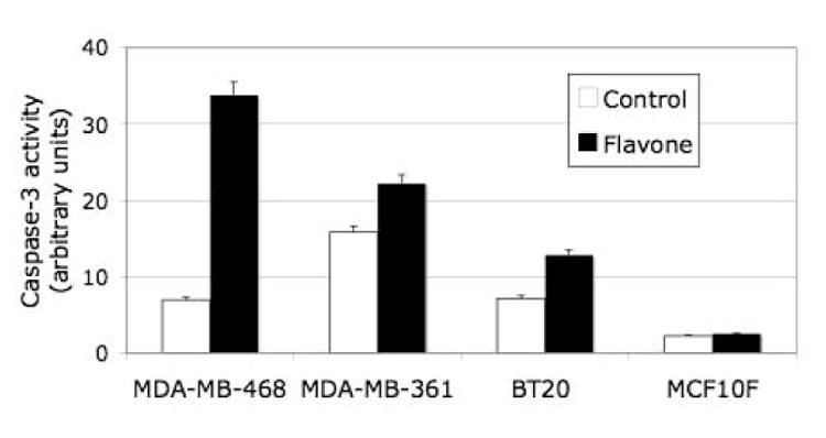
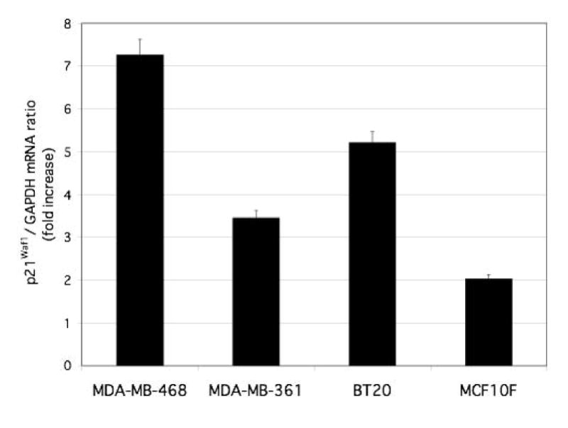
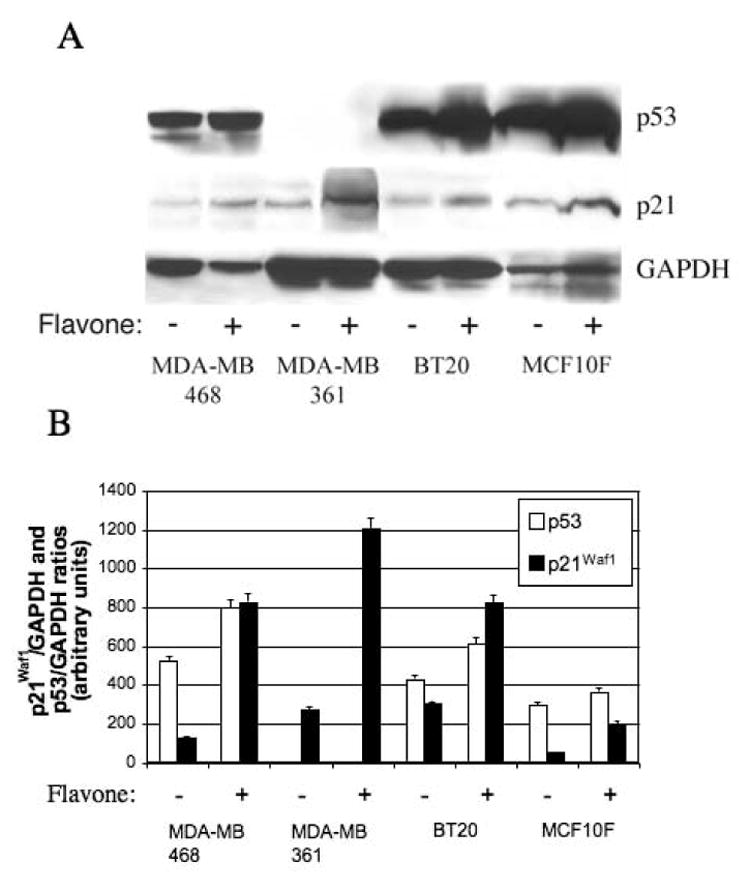
Results: We show that exposure to 150 microM flavone increased DLC1 expression in breast but not in liver or prostate carcinoma cells or a nonmalignant breast epithelial cell line. Flavone restored the expression of DLC1 in the breast carcinoma cell lines MDA-MB-468, MDA-MB-361, and BT20 as well as in the colon carcinoma cell line HT-29 all of which are DLC-1-negative due to promoter hypermethylation. We further show that flavone inhibited cell proliferation, induced cell cycle arrest at G(2)-M, increased p21(Waf1) gene expression, and caused apoptosis. Microarray analysis of these aggressive and metastatic breast carcinoma cells revealed 29 flavone-responsive genes, among which the DNA damage-inducible GADD genes were up-regulated and the proto-oncogene STMN1 and IGFBP3 were down-regulated.

Conclusions: Flavone-mediated alterations of genes that regulate tumor cell proliferation, cell cycle, and apoptosis contribute to chemopreventive and antitumoral effects of flavone. Alone or in combination with demethylating agents, flavone may be an effective adjunct to chemotherapy in preventing breast cancer metastasis.


Figures

Figure 1
Figure 1
Effects of flavone on DLC1 expression in HT-29 colon carcinoma cells, breast carcinoma cells (MDA-MB-468, MDA-MB-361, BT20), and a normal breast epithelial cell line (MCF10F). Cells were incubated in the absence or presence of 150 μM flavone for 24 h, after which total RNA was isolated and subjected to quantitative RT-PCR analysis of DLC-1 mRNA. The level of normalized DLC1 expression for flavone-treated cells is shown relative to that in nontreated cells (see materials and methods). Data are the means ± SEM from independent experiments.
Figure 2
Figure 2
Inhibition of cell proliferation by flavone in breast carcinoma cell lines and a normal breast epithelial cell line. Cells were incubated in the absence or presence of 150 μM flavone for the indicated times, after which cell number was determined by the MTT assay. Cell number in flavone-treated cultures was expressed as a percentage of that in control cultures. Data are means ± SD of triplicates from a representative experiment.
Figure 3
Figure 3
Effect of flavone on cell cycle distribution in breast carcinoma cell lines and a nonmalignant breast epithelial cell line. Cells were incubated in the absence (control) or presence of 150μM flavone for 24 h, after which DNA content was determined by flow cytometry. Arrows indicate the sub-G1 population, and the percentages of cells in G0-G1 or G2-M are shown.
Figure 4
Figure 4
Effect of flavone on caspase-3 activity in breast carcinoma cell and nonmalignant breast epithelial cells. Cells were incubated in the absence or presence of 150μM flavone for 24 h, after which the activity of caspase-3 was determined. Data are expressed in arbitrary units and are means ± SD of triplicates from a representative experiment.
Figure 5
Figure 5
Effect of flavone on p21Waf1 gene expression in breast carcinoma cells and nonmalignant breast epithelial cells. Cells were incubated in the absence or presence of 150 μM flavone for 24 h, after which total RNA was isolated and subjected to quantitative RT-PCR analysis of p21Waf1 mRNA. Data were normalized by the amount of GAPDH mRNA, and the effect of flavone was evaluated by the 2−ΔΔct method and considered significant when 2−ΔΔct was ≥2 or ≤0.5. The abundance of p21Waf1 mRNA in flavone-treated cells is shown relative to that in nontreated cells. Data are the means ± SEM from independent experiments.
Figure 6
Figure 6
Effects of flavone on p21Waf1 and p53 abundance in breast cancer cell lines) and a normal breast epithelial cell line. (A) Cells were incubated in the absence or presence of 150μM flavone for 24 h, after which cell lysates were subjected to immunoblot analysis with antibodies to p53, to p21Waf1, or to GAPDH. (B) Blots similar to that in (A) were subjected to densitometric analysis, and the amounts of p21Waf1 and p53 were normalized by that of GAPDH.
Figure 7
Figure 7
Effect of ectopic DLC-1 expression on p21Waf1 abundance in the breast cancer cell lines MDA-MB-468, MDA-MB-361, and BT20. Cells were transfected with expression vectors for DLC-1 or LacZ (control), and the abundance of DLC-1 and p21Waf1 mRNAs was subsequently determined by quantitative RT-PCR analysis. Data were normalized by the amount of GAPDH mRNA, and the normalized data were expressed relative to the corresponding value for cells transfected with the control vector. Differences between cells transfected with the DLC-1 vector and those transfected with the control vector were evaluated by the 2−ΔΔct method and considered significant when 2−ΔΔct was ≥2 or ≤0.5. Data are the means ± SEM from independent experiments.

Similar articles

Cited by

References

    1. Yuan BZ, Miller MJ, Keck CL, Zimonjic DB, Thorgeirsson SS, Popescu NC. Cloning, characterization, and chromosomal localization of a gene frequently deleted in human liver cancer (DLC-1) homologous to rat RhoGAP. Cancer Res. 1998;58:2196–99. - PubMed
    1. Goodison S, Yuan J, Sloan D, Kim R, Li C, Popescu NC, et al. The RhoGAP protein DLC-1 functions as a metastasis suppressor in breast cancer cells. Cancer Res. 2005;65:6042–53. - PMC - PubMed
    1. Seng TJ, Low JS, Li H, Cui Y, Goh HK, Wong ML, et al. The major 8p22 tumor suppressor DLC1 is frequently silenced by methylation in both endemic and sporadic nasopharyngeal, esophageal, and cervical carcinomas, and inhibits tumor cell colony formation. Oncogene advance online publication. 2006 July 24; doi: 10.1038/sj.onc.1209839. - DOI - PubMed
    1. Syed V, Mukherjee K, Lyons-Weiler J, Lau KM, Mashima T, Tsuruo T, et al. Identification of ATF-3, caveolin-1, DLC-1, and NM23-H2 as putative antitumorigenic, progesterone-regulated genes for ovarian cancer cells by gene profiling. Oncogene. 2005;24:1774–87. - PubMed
    1. Wong CM, Yam JW, Ching YP, Yau TO, Leung TH, Jin DY, et al. Rho GTPase-activating protein deleted in liver cancer suppresses cell proliferation and invasion in hepatocellular carcinoma. Cancer Res. 2005;65:8861–68. - PubMed

Publication types

MeSH terms