. 2007 May 2;129(17):5558-69.
doi: 10.1021/ja068511u. Epub 2007 Apr 10.

Macrocyclic beta-sheet peptides that mimic protein quaternary structure through intermolecular beta-sheet interactions

Affiliations

Macrocyclic beta-sheet peptides that mimic protein quaternary structure through intermolecular beta-sheet interactions

Omid Khakshoor et al. J Am Chem Soc. .

Abstract

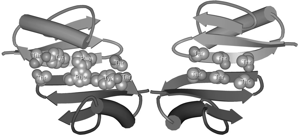
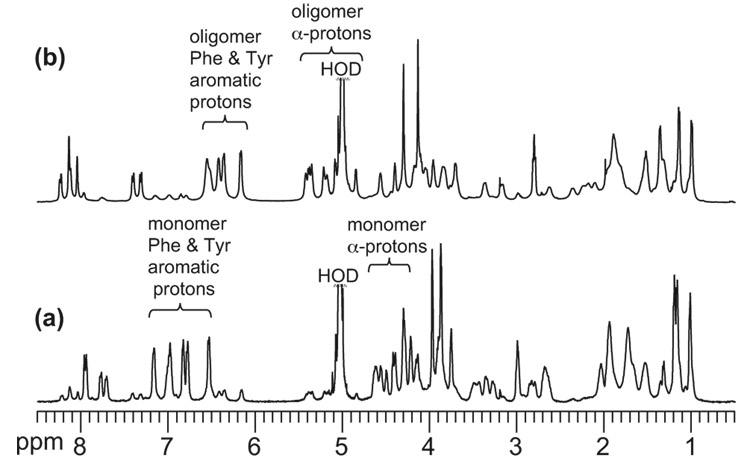
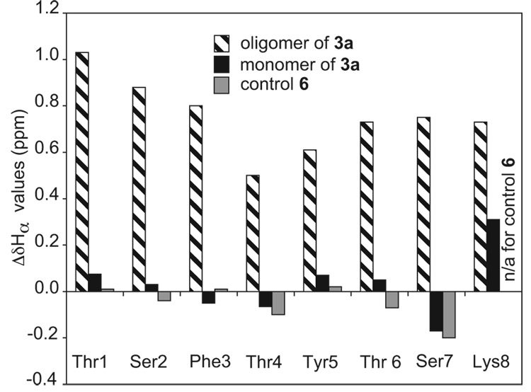
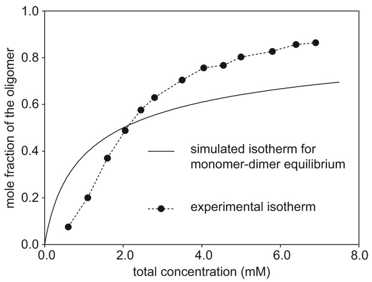
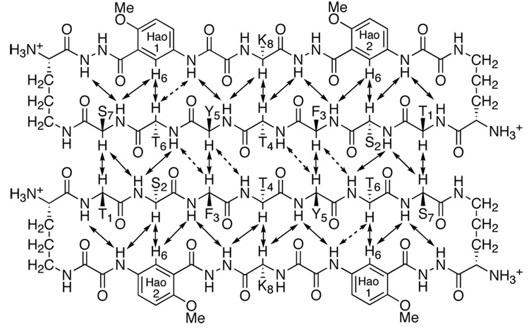
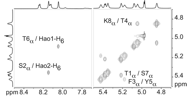
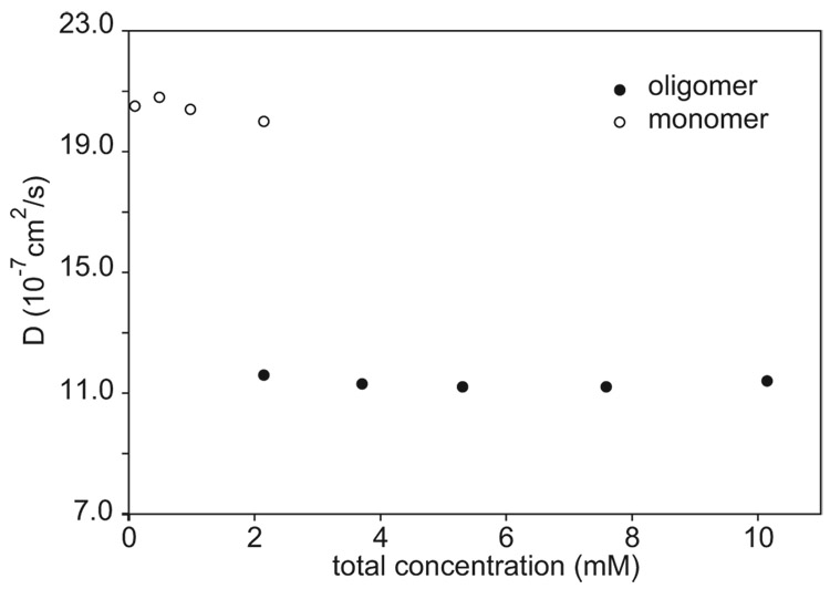
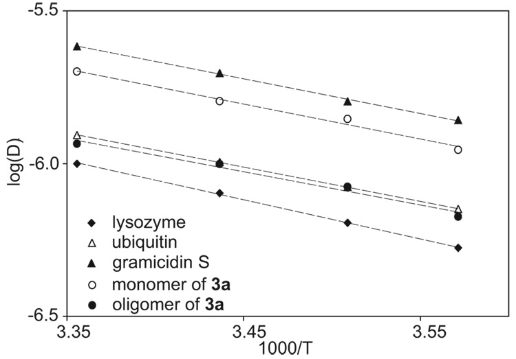
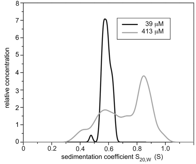
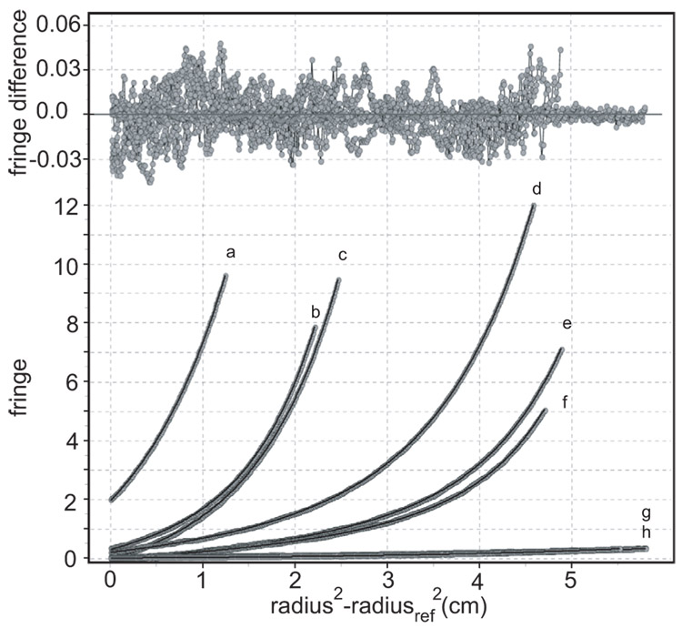
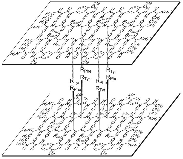
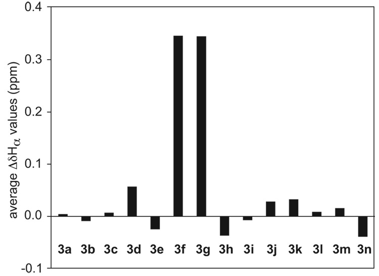
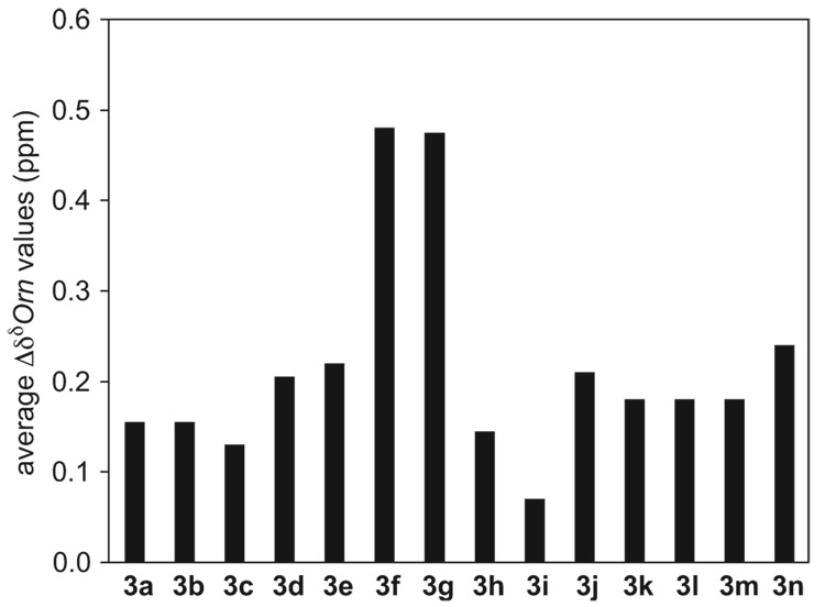
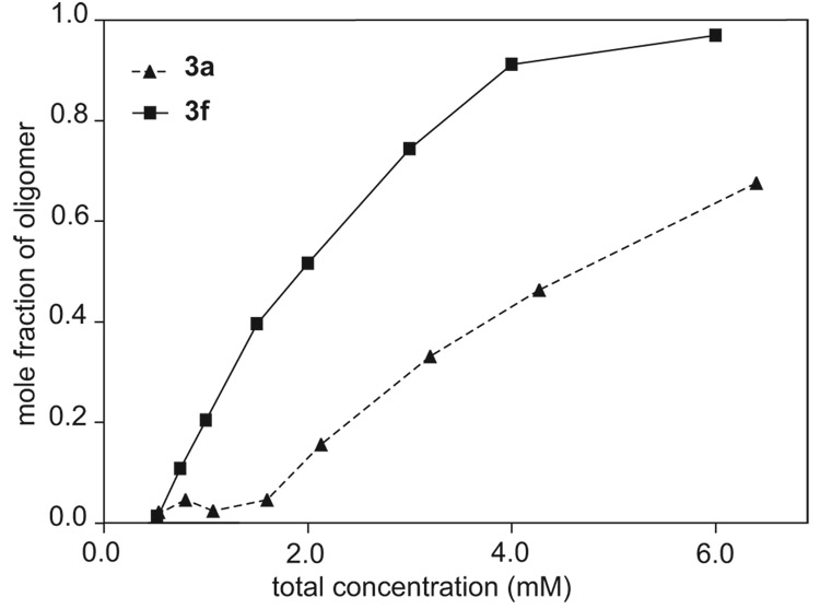
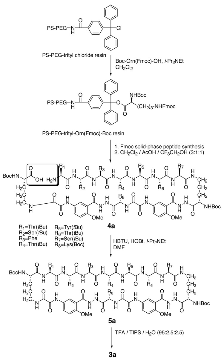
This paper reports the design, synthesis, and characterization of a family of cyclic peptides that mimic protein quaternary structure through beta-sheet interactions. These peptides are 54-membered-ring macrocycles comprising an extended heptapeptide beta-strand, two Hao beta-strand mimics [JACS 2000, 122, 7654] joined by one additional alpha-amino acid, and two delta-linked ornithine beta-turn mimics [JACS 2003, 125, 876]. Peptide 3a, as the representative of these cyclic peptides, contains a heptapeptide sequence (TSFTYTS) adapted from the dimerization interface of protein NuG2 [PDB ID: 1mio]. 1H NMR studies of aqueous solutions of peptide 3a show a partially folded monomer in slow exchange with a strongly folded oligomer. NOE studies clearly show that the peptide self-associates through edge-to-edge beta-sheet dimerization. Pulsed-field gradient (PFG) NMR diffusion coefficient measurements and analytical ultracentrifugation (AUC) studies establish that the oligomer is a tetramer. Collectively, these experiments suggest a model in which cyclic peptide 3a oligomerizes to form a dimer of beta-sheet dimers. In this tetrameric beta-sheet sandwich, the macrocyclic peptide 3a is folded to form a beta-sheet, the beta-sheet is dimerized through edge-to-edge interactions, and this dimer is further dimerized through hydrophobic face-to-face interactions involving the Phe and Tyr groups. Further studies of peptides 3b-3n, which are homologues of peptide 3a with 1-6 variations in the heptapeptide sequence, elucidate the importance of the heptapeptide sequence in the folding and oligomerization of this family of cyclic peptides. Studies of peptides 3b-3g show that aromatic residues across from Hao improve folding of the peptide, while studies of peptides 3h-3n indicate that hydrophobic residues at positions R3 and R5 of the heptapeptide sequence are important in oligomerization.


Figures

Figure 1
Figure 1
Views from the front and back sides of the crystallographic NuG2 dimer illustrating the heptapeptide β-strand dimerization interface TTFTYTT (PDB ID: 1mio).
Figure 2
Figure 2
1H NMR spectra of peptide 3a at concentrations of 1.0 mM (a) and 6.0 mM (b) in D2O at 500 MHz and 280 K.
Figure 3
Figure 3
Deviation of the α-proton chemical shifts from published random coil values (ΔδHα = observed δHα - random coil δHα) for the oligomer, the monomer, and control peptide 6. 1H NMR studies were performed on 8.5 mM and 1.0 mM solutions of peptide 3a and on an 8.0 mM solution of control peptide 6 in D2O at 280 K.
Figure 4
Figure 4
Concentration dependence of oligomer formation of peptide 3a: mole fraction of oligomer vs. total peptide concentration. The relative concentrations of the monomer and oligomer were determined in D2O at 280 K by integrating the corresponding 1H NMR resonances. A simulated isotherm for a monomer-dimer equilibrium (K1,2 = 500 M−1), which is shown for comparison, does not fit the data.
Figure 5
Figure 5
Key NOEs involving main-chain inter-residue contacts in the β-sheet dimer of peptide 3a. Dashed double-headed arrows represent weak or ambiguous NOEs.
Figure 6
Figure 6
Selected expansions of the NOESY spectrum of the peptide 3a oligomer in D2O illustrating key NOEs resulting from folding and edge-to-edge dimerization in the oligomer. NOESY studies of an 8.5 mM solution of peptide 3a were performed at 800 MHz and 280 K with a 75-ms mixing time.
Figure 7
Figure 7
The diffusion coefficients of the oligomer and the monomer of peptide 3a at various total concentrations of the peptide. The diffusion coefficients were measured in D2O at 298 K by using an sLED pulse sequence on an 800 MHz 1H NMR spectrometer.
Figure 8
Figure 8
Comparison of the temperature dependence of the diffusion coefficients of the monomer and the oligomer of peptide 3a to those of lysozyme, ubiquitin, and gramicidin S in D2O. The diffusion coefficients were measured at 280, 285, 291, and 298 K by using an sLED pulse sequence on an 800 MHz 1H NMR spectrometer. The diffusion coefficients of these species were measured at the following concentrations: 3a monomer, 2 mM; 3a oligomer, 10 mM; lysozyme, 1.4 mM; ubiquitin, 1.0 mM; and gramicidin S, 1.1 mM.
Figure 9
Figure 9
Van Holde–Weischet distributions of sedimentation velocity experiments of peptide 3a measured with UV absorbance at 280 nm at 39 µM (black line) and with Rayleigh interference at 413 µM (grey line).
Figure 10
Figure 10
Sedimentation equilibrium data for peptide 3a fitted to a monomer-tetramer equilibrium model. Residuals of the fit are shown on the top, overlays on the bottom. Grey points represent experimental data at (a) 9 mM and 60 000 rpm, (b) 9 mM and 55 000 rpm, (c) 9 mM and 50 000 rpm, (d) 1.5 mM and 60 000 rpm, (e) 1.5 mM and 55 000 rpm, (f) 1.5 mM and 50 000 rpm, (g) 15 µM and 60 000 rpm, and (h) 15 µM and 55 000 rpm. Black curves represent the fitted model.
Figure 11
Figure 11
Illustration of the β-sheet sandwich tetramer of peptide 3a.
Figure 12
Figure 12
Average ΔδHα values of the residues at positions R1–R7 for peptide 3a3n monomers at 298 K in D2O.
Figure 13
Figure 13
Average ΔδδOrn values for peptide 3a3n monomers at 298 K in D2O.
Figure 14
Figure 14
Comparison of the concentration-dependent oligomerization of peptides 3a and 3f: mole fraction of oligomer vs. total peptide concentration. The relative concentrations of the monomer and oligomer were determined in D2O at 298 K by integrating the corresponding 1H NMR resonances.
Scheme 1
Scheme 1
Synthesis of peptide 3a

References

    1. Maitra S, Nowick JS. In: The Amide Linkage: Structural Significance in Chemistry, Biochemistry, and Materials Science. Greenberg A, Breneman CM, Liebman JF, editors. New York: Wiley; 2000. pp. 495–518.
    2. Remaut H, Waksman G. Trends Biochem. Sci. 2006;31:436–444. - PubMed
    1. Navia MA, Fitzgerald PMD, McKeever BM, Le CT, Heimbach JC, Herber WK, Sigal IS, Darke PL, Springer JP. Nature. 1989;337:615–620. - PubMed
    1. Nassar N, Horn G, Herrmann C, Scherer A, McCormick F, Wittinghofer A. Nature. 1995;375:554–560. - PubMed
    1. Ross CA, Poirier MA. Nat. Med. 2004;10 Suppl.:S10–S17. - PubMed
    2. Tycko R. Curr. Opin. Struct. Biol. 2004;14:96–103. - PubMed
    3. Makin OS, Serpell LC. FEBS J. 2005;272:5950–5961. - PubMed
    4. Nelson R, Eisenberg D. Curr. Opin. Struct. Biol. 2006;16:260–265. - PubMed
    5. Chiti F, Dobson CM. Annu. Rev. Biochem. 2006;75:333–366. - PubMed
    6. Wetzel R. Acc. Chem. Res. 2006;39:671–679. - PubMed
    1. Sommers WS, Phillips SEV. Nature. 1992;359:387–393. - PubMed

Publication types