Review
. 2007:97:275-94.
doi: 10.1016/S0065-230X(06)97012-2.

Extracellular matrix, nuclear and chromatin structure, and gene expression in normal tissues and malignant tumors: a work in progress

Affiliations
Review

Extracellular matrix, nuclear and chromatin structure, and gene expression in normal tissues and malignant tumors: a work in progress

Virginia A Spencer et al. Adv Cancer Res. 2007.

Abstract

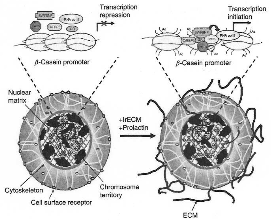
Almost three decades ago, we presented a model where the extracellular matrix (ECM) was postulated to influence gene expression and tissue-specificity through the action of ECM receptors and the cytoskeleton. This hypothesis implied that ECM molecules could signal to the nucleus and that the unit of function in higher organisms was not the cell alone, but the cell plus its microenvironment. We now know that ECM invokes changes in tissue and organ architecture and that tissue, cell, nuclear, and chromatin structure are changed profoundly as a result of and during malignant progression. Whereas some evidence has been generated for a link between ECM-induced alterations in tissue architecture and changes in both nuclear and chromatin organization, the manner by which these changes actively induce or repress gene expression in normal and malignant cells is a topic in need of further attention. Here, we will discuss some key findings that may provide insights into mechanisms through which ECM could influence gene transcription and how tumor cells acquire the ability to overcome these levels of control.


Figures

Fig. 1
Fig. 1
A schematic diagram illustrating the basic principles of dynamic reciprocity between neighboring cells and their extracellular environment. Mechanical and biochemical signals received at points of cell–cell or cell–ECM contact are transduced to the nucleus by transmembrane receptors, signaling molecules, and cytoskeletal components where they initiate nuclear events resulting in the expression of specific gene products that are excreted back into the extracellular milieu. Green arrows represent the bidirectional flow of mechanical and biochemical signals between the ECM and the nucleus. RTKs represent receptor tyrosine kinase. (Modified, with permission, from Bissell et al., 2005.) (See Color Insert.)
Fig. 2
Fig. 2
An illustration of the different levels through which ECM controls gene expression and tissue function. As cells transition from a 2D monolayer to a 3D environment, they undergo changes in cell shape that influence the expression of certain genes. Exposure to ECM engages specific cell surface receptors and initiates the transduction of biochemical and mechanical signals through the cell to the nucleus, where they further influence gene expression. As the duration of exposure time to ECM increases, cells undergo morphogenic events involving the formation of acinar structures and once again exhibit changes in their gene expression profile. Thus, tissue structure influences gene expression and, therefore, dictates tissue function. (Modified, with permission, from Bissell et al., 1999, 2005; Roskelley et al., 1995.) (See Color Insert.)
Fig. 3
Fig. 3
Schematic representation of lrECM- and prolactin-induced changes in nuclear organization and transcription factor binding in mammary epithelial cells that lead to the initiation of β-casein gene transcription. In the absence of prolactin and lrECM, STAT5 is predominantly cytoplasmic in primary cells. Exposure to lrECM and prolactin induces STAT5 phosphorylation, nuclear translocation, and binding to its cognate DNA sequence in the β-casein promoter. In addition, this treatment increases the association of acetylated histones and promotes the binding of additional transcription factors including C/EBPβ, SWI/SNF, GR, and RNA polymerase (pol) II to the β-casein promoter. Ac represents acetylation at lysine residues along histone N-terminal tails. (See Color Insert.)

References

    1. Anders M, Hansen R, Ding RX, Rauen KA, Bissell MJ, Korn WM. Disruption of 3D tissue integrity facilitates adenovirus infection by deregulating the cox-sackievirus and adenovirus receptor. Proc. Natl. Acad. Sci. USA. 2003;100:1943–1948. - PMC - PubMed
    1. Barcellos-Hoff MH, Aggeler J, Ram TG, Bissell MJ. Functional differentiation and alveolar morphogenesis of primary mammary cultures on reconstituted basement membrane. Development. 1989;105:223–235. - PMC - PubMed
    1. Baskaran R, Dahmus ME, Wang JY. Tyrosine phosphorylation of mammalian RNA polymerase II carboxyl-terminal domain. Proc. Natl. Acad. Sci. USA. 1993;90:11167–11171. - PMC - PubMed
    1. Baskaran R, Chiang GG, Wang JY. Identification of a binding site in c-Ab1 tyrosine kinase for the C-terminal repeated domain of RNA polymerase II. Mol. Cell. Biol. 1996;16:3361–3369. - PMC - PubMed
    1. Biggiogera M, Bottone MG, Scovassi AI, Soldani C, Vecchio L, Pellicciari C. Rearrangement of nuclear ribonucleoprotein (RNP)-containing structures during apoptosis and transcriptional arrest. Biol. Cell. 2004;96:603–615. - PubMed

Publication types

MeSH terms