. 2007 Apr;12(4):515-29.
doi: 10.1016/j.devcel.2007.01.019.

A microtubule-independent role for centrosomes and aurora a in nuclear envelope breakdown

Affiliations

A microtubule-independent role for centrosomes and aurora a in nuclear envelope breakdown

Nathan Portier et al. Dev Cell. 2007 Apr.

Abstract

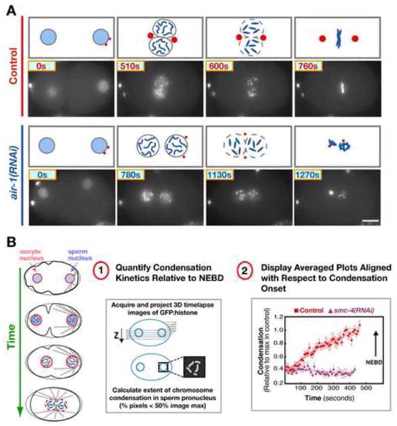
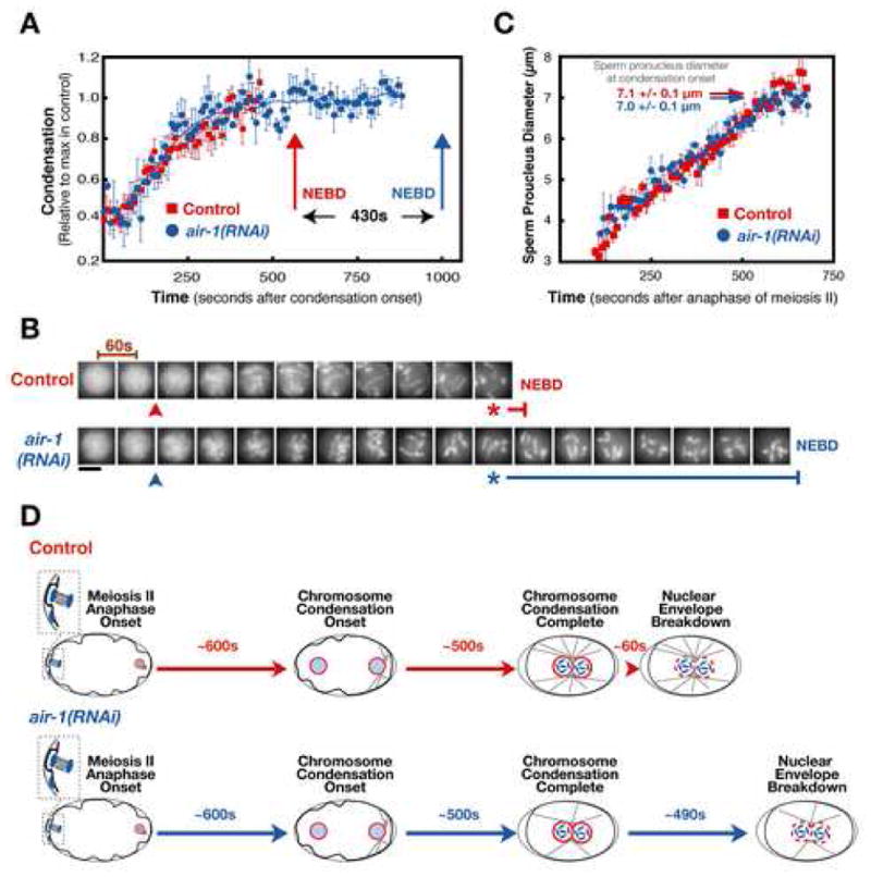
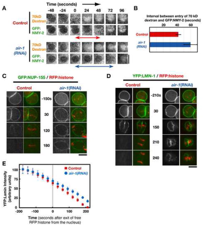
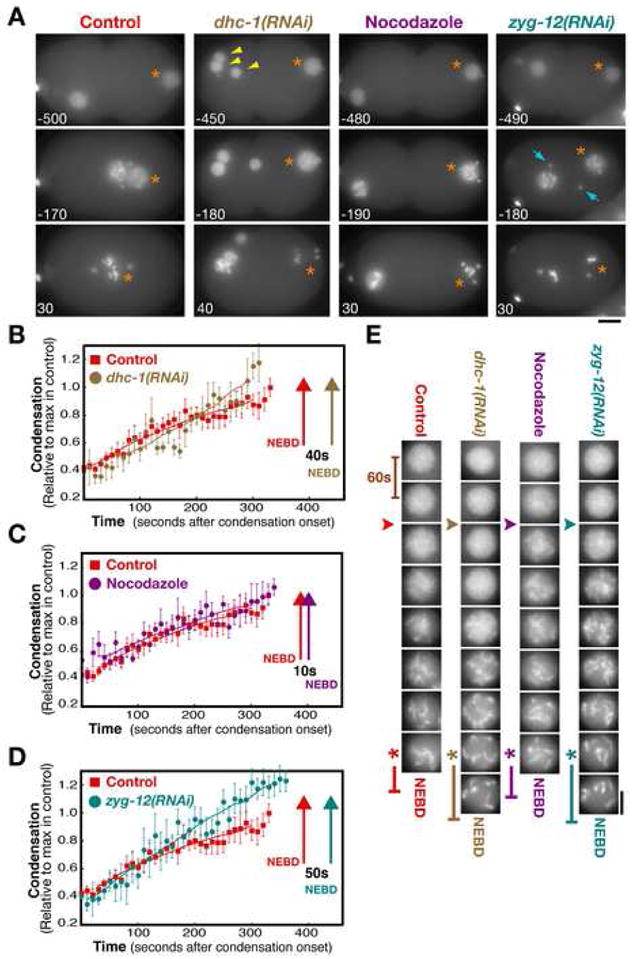
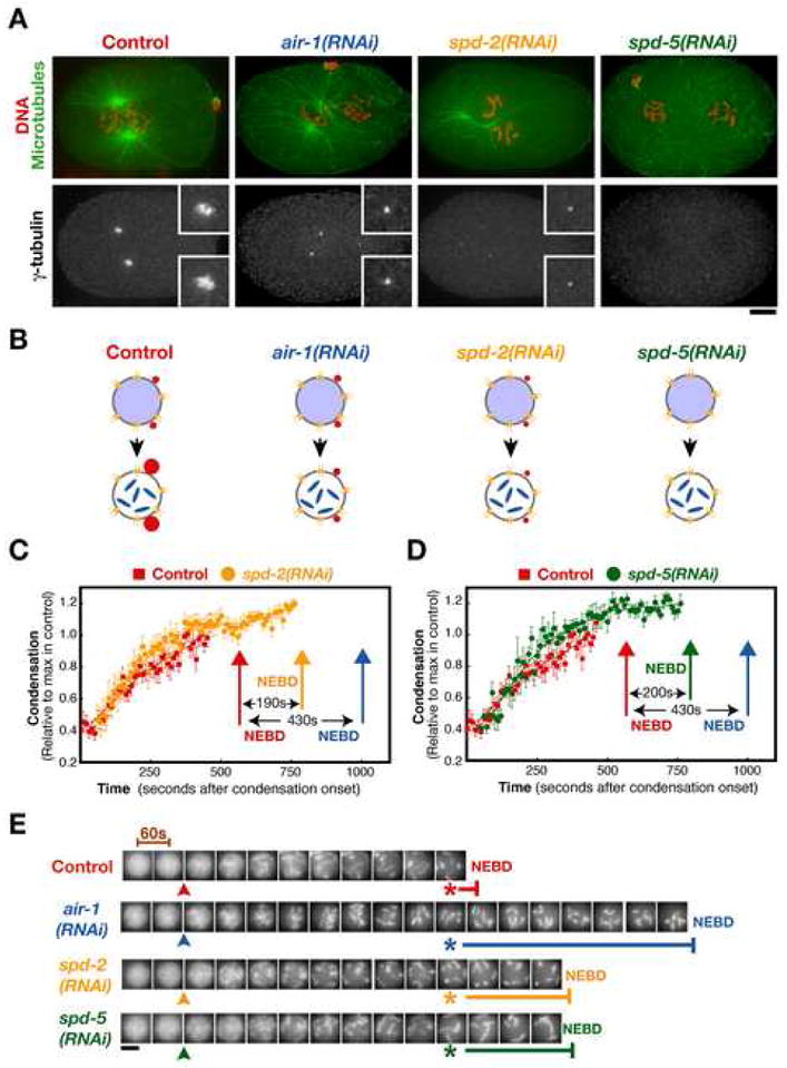
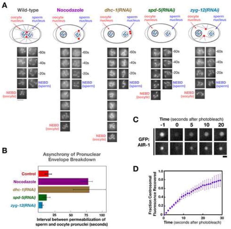
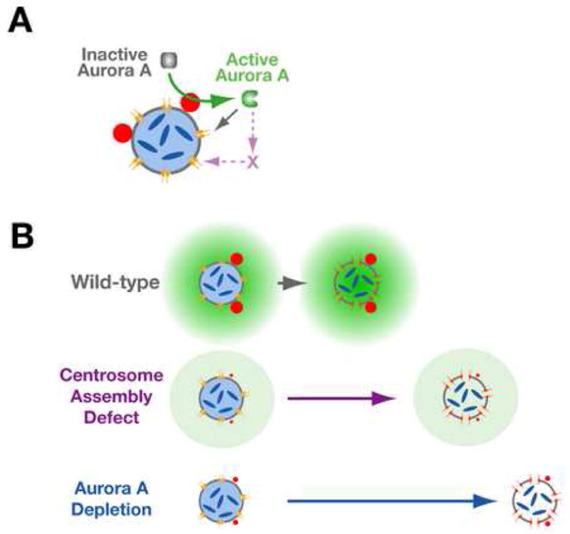
Aurora A kinase localizes to centrosomes and is required for centrosome maturation and spindle assembly. Here we describe a microtubule-independent role for Aurora A and centrosomes in nuclear envelope breakdown (NEBD) during the first mitotic division of the C. elegans embryo. Aurora A depletion does not alter the onset or kinetics of chromosome condensation, but dramatically lengthens the interval between the completion of condensation and NEBD. Inhibiting centrosome assembly by other means also lengthens this interval, albeit to a lesser extent than Aurora A depletion. By contrast, centrosomally nucleated microtubules and the nuclear envelope-associated motor dynein are not required for timely NEBD. These results indicate that mitotic centrosomes generate a diffusible factor, which we propose is activated Aurora A, that promotes NEBD. A positive feedback loop, in which an Aurora A-dependent increase in centrosome size promotes Aurora A activation, may temporally couple centrosome maturation to NEBD during mitotic entry.


Figures

Figure 1
Figure 1. Aurora A Is Required for Centrosome Maturation and for Proper Timing of Events During Mitotic Entry
(A) Selected stills from time-lapse sequences of control (top) and aurora A depleted (air-1(RNAi); bottom) embryos expressing GFP:histone. Times are seconds after the first panel of each sequence. Schematics above each panel also illustrate the previously characterized (Hannak et al., 2001) effect of aurora A depletion on centrosome (red) maturation. Bar, 10 μm. (B) Schematics illustrate the stages between the onset of chromosome condensation and NEBD of the first mitotic division that follows fertilization. (B-1) Outline of the image analysis method (Maddox et al., 2006) used to quantitatively monitor the kinetics of chromosome condensation. (B-2) Plot of the averaged condensation parameter versus time for control (red squares; n=12 embryos) and smc-4(RNAi) (purple triangles; n=8 embryos) embryos. The average value of the condensation parameter was calculated after aligning the sequences with respect to NEBD. Traces are displayed with the onset of condensation in controls as t=0 (arrow marks NEBD in both data sets). Error bars are SEM.
Figure 2
Figure 2. Depletion of Aurora A Specifically Delays Nuclear Envelope Breakdown
Aurora A depleted embryos condense chromosomes with normal kinetics but exhibit a dramatic delay between the completion of condensation and NEBD. (A) Plot of the average of the condensation parameter versus time for control (red squares; n=12) and aurora A depleted embryos (air-1(RNAi); blue circles; n=8). The average value of the condensation parameter was calculated after aligning the sequences with respect to NEBD. Traces are displayed aligned with respect to the onset of condensation. Error bars are SEM. (B) Representative images of the sperm pronucleus in a control and an aurora A depleted embryo. Images are at 60s intervals. The onset (arrowheads) and completion (asterisks) of condensation defined by the analysis in (A) are indicated, as is the timing of NEBD (vertical bars). Scale bar is 5 μm. (C) Plot of the average diameter of the sperm pronucleus vs. time after the onset of anaphase of meiosis II in control (red squares; n=5) and aurora A depleted (air-1(RNAi); blue circles; n=5) embryos. The average pronuclear diameter at the onset of condensation calculated from the data in (A) is indicated for control (red arrow) and aurora A depleted (blue arrow) embryos. (D) Schematic summarizing the timing of events during the first mitotic division in control and aurora A-depleted embryos. Chromosomes initiate and complete condensation with identical timing relative to anaphase of meiosis II in both types of embryos. However, nuclear envelope permeabilization is dramatically delayed by aurora A depletion.
Figure 3
Figure 3. Aurora A Depletion Slows the Progression of Nuclear Envelope Breakdown in Addition to Dramatically Delaying its Onset
(A) Selected stills from time-lapse sequences of nuclei in control and aurora A depleted embryos that are expressing GFP:NMY-2 and contain Texas Red conjugated 70kD dextran introduced by injection. Images are shown at 24s intervals. Times are in seconds relative to entry of 70kD dextran into the nuclear space. (B) Bar graph of the average interval between the entry of 70kD dextran and GFP:NMY-2 into the nuclear space (indicated by the double-headed arrows in A, measured in 10 control and 7 air-1(RNAi) embryos. Error bars are the 95% confidence interval. (C,D) Selected stills from time-lapse sequences of nuclei in control and aurora A depleted embryos co-expressing RFPmCherry:histone and either GFP:NUP-155 (C) or YFP:LMN-1 (D). Times are in seconds relative to exit of free RFP:histone from the nucleus. Bars, 5 μm. (E) Graph plotting the average intensity of YFP:LMN-1 associated with the nuclear periphery as a function of time in seconds relative to loss of free RFP:histone from the nucleus (n=6 for each condition; error bars are the 80% confidence interval).
Figure 4
Figure 4. Centrosomal Microtubules and Nuclear Envelope-Associated Dynein are Not Critical for the Timing of Nuclear Envelope Permeabilization
(A) Selected stills from time-lapse sequences of control, dynein depleted (dhc-1(RNAi)), nocodozole treated, and zyg-12(RNAi) embryos expressing GFP:histone and GFP:γ-tubulin. The sperm pronucleus (orange asterisks), extra maternal pronuclei resulting from failure of meiotic segregation in the DHC-1 depleted embryo (yellow arrowheads), and the centrosomes in the ZYG-12 depleted embryo (blue arrows) are indicated. Times are with respect to the exit of free GFP histone from the nucleus. Bar, 10μm. (B–D) Plots comparing condensation kinetics in control embryos (n=10; red squares) with those in dhc-1(RNAi) (B; brown circles, n=6), nocodazole treated (C; purple circles, n=7), and zyg-12(RNAi) (D; cyan circles, n=10) embryos. Arrows mark the timing of NEBD for each condition. Error bars are SEM. (E) Representative images of the sperm pronucleus in a control, a dhc-1(RNAi), a nocodazole-treated, and a zyg-12(RNAi) embryo. Images are at 60s intervals. The onset (arrowheads) and completion (asterisks) of condensation defined by the analysis in (B–D) are indicated, as is the timing of NEBD (vertical bars). Bar, 5 μm.
Figure 5
Figure 5. Inhibiting Centrosome Assembly Delays Nuclear Envelope Breakdown, but to a Lesser Extent than Depletion of Aurora A
(A) Prophase control, air-1(RNAi), spd-2(RNAi) and spd-5(RNAi) embryos just prior to or after NEBD were fixed and stained for DNA and microtubules (red and green; top panels) and γ-tubulin (lower panels). Bar, 5 μm. Insets are magnified 2.5-fold. (B) Schematic illustrating the consequences of depleting each of the indicated proteins on centrosome structure. (C,D) Plots of the condensation parameter versus time comparing control (n=12) and SPD-2 depleted (C; n=10), or SPD-5 depleted (D; n=6), embryos. The timing of NEBD in control (red arrows), SPD-2 depleted (yellow arrow), and SPD-5 depleted (green arrow) embryos is indicated. For comparison, the timing of NEBD in embryos depleted of aurora A (blue arrows; replotted from Fig. 2) is also shown. Error bars are SEM. (E) Representative images of the sperm pronucleus from a control, an air-1(RNAi), a spd-2(RNAi) and a spd-5(RNAi) embryo. Images are at 60s intervals. The onset (arrowheads) and completion (asterisks) of condensation defined by the analysis in (C, D) are indicated, as is the timing of NEBD (horizontal bars). Scale bar is 5 μm.
Figure 6
Figure 6. Centrosomes Generate a Diffusible Factor that Promotes Nuclear Envelope Permeabilization
(A) Images of the oocyte and sperm pronuclei in representative embryos for each condition. Schematics above each set of images illustrate the effect of the perturbations on the relative positions of the centrosomes and nuclei. (B) Graph of the interval between permeabilization of the oocyte and sperm-derived pronuclei to GFP:histone (time of oocyte pronuclear permeabilization - time of sperm pronuclear permeabilization) in control embryos, and embryos that fail to undergo pronuclear migration. In contrast to control embryos (n=12), permeabilization of the oocyte and sperm derived pronuclei is asynchronous in nocodazole-treated (n=5) and dynein depleted (dhc-1(RNAi); n=5) embryos, with the oocyte pronucleus breaking down later than the sperm pronucleus. This asynchrony is not observed in embryos in which pronuclear migration fails due to lack of functional centrosomes (spd-5(RNAi); n=5) or when centrosomes do not remain in proximity to the sperm pronucleus (zyg-12(RNAi); n=10). Error bars are the 80% confidence interval. (C) Selected still images from two representative experiments in which the recovery of centrosomal GFP:AIR-1 fluorescence was monitored after photobleaching. Times in seconds after the bleach. Bar, 1 μm. (D) Graph shows the average fraction of GFP fluorescence recovered as a function of time in seconds after the photobleach (n=8; error bars are the 80% confidence interval).
Figure 7
Figure 7. Model for the Role of Aurora A in Coordinating Centrosome Maturation with NEBD
(A) We propose that the mitotic centrosome scaffold harbors an aurora A activator. Activated aurora A promotes NEBD, the first step of which is permeabilization of the envelope due to loss of peripheral components of the nuclear pores (Lenart et al., 2003; Terasaki et al., 2001). The role of aurora A in pore opening could be via direct phosphorylation of pore components or mediated by as yet unknown intermediates. (B) In wild-type embryos, a positive feedback loop in which the aurora A-dependent increase in centrosome size increases the rate of aurora A activation, generates a gradient of active aurora A with the maximum concentration near the centrosomes. When centrosomes are mature and the local concentration of active aurora A is sufficiently high, the nuclear envelope becomes permeable. In the absence of centrosomes, the activation of aurora A and permeabilization of the nuclear envelope are delayed. In aurora A depleted embryos an even greater delay is observed.

Comment in

References

    1. Andrews PD. Aurora kinases: shining lights on the therapeutic horizon? Oncogene. 2005;24:5005–5015. - PubMed
    1. Audhya A, Hyndman F, McLeod IX, Maddox AS, Yates JR, 3rd, Desai A, Oegema K. A complex containing the Sm protein CAR-1 and the RNA helicase CGH-1 is required for embryonic cytokinesis in Caenorhabditis elegans. J Cell Biol. 2005;171:267–79. - PMC - PubMed
    1. Bar-Shira A, Pinthus JH, Rozovsky U, Goldstein M, Sellers WR, Yaron Y, Eshhar Z, Orr-Urtreger A. Multiple genes in human 20q13 chromosomal region are involved in an advanced prostate cancer xenograft. Cancer Res. 2002;62:6803–6807. - PubMed
    1. Barr FA, Sillje HH, Nigg EA. Polo-like kinases and the orchestration of cell division. Nat Rev Mol Cell Biol. 2004;5:429–440. - PubMed
    1. Beaudouin J, Gerlich D, Daigle N, Eils R, Ellenberg J. Nuclear envelope breakdown proceeds by microtubule-induced tearing of the lamina. Cell. 2002;108:83–96. - PubMed

Publication types

MeSH terms