. 2007 Jun 8;282(23):17061-8.
doi: 10.1074/jbc.M700490200. Epub 2007 Apr 9.

Nucleoplasmic calcium is required for cell proliferation

Affiliations

Nucleoplasmic calcium is required for cell proliferation

Michele A Rodrigues et al. J Biol Chem. .

Abstract

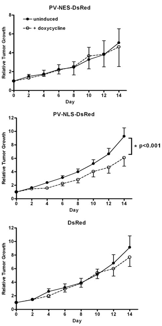
Ca(2+) signals regulate cell proliferation, but the spatial and temporal specificity of these signals is unknown. Here we use selective buffers of nucleoplasmic or cytoplasmic Ca(2+) to determine that cell proliferation depends upon Ca(2+) signals within the nucleus rather than in the cytoplasm. Nuclear Ca(2+) signals stimulate cell growth rather than inhibit apoptosis and specifically permit cells to advance through early prophase. Selective buffering of nuclear but not cytoplasmic Ca(2+) signals also impairs growth of tumors in vivo. These findings reveal a major physiological and potential pathophysiological role for nucleoplasmic Ca(2+) signals and suggest that this information can be used to design novel therapeutic strategies to regulate conditions of abnormal cell growth.


Figures

FIGURE 1
FIGURE 1. Adenoviral delivery of the Ca2+ buffer protein PV targeted to the nucleus or cytoplasm
A, subcellular localization of targeted PV constructs. NLS or NES sequences are used to target PV to the nucleus or cytoplasm, respectively, and each construct is tagged with DsRed to monitor expression and subcellular location. An adenoviral delivery system is used for each construct. Confocal microscopy demonstrates very high transfection rates and that PV-NLS-DsRed is expressed in the nucleus, PV-NES-DsRed is expressed in the cytoplasm, and DsRed alone, which serves as a transfection control, is expressed in both compartments. In each image, red indicates DsRed, blue indicates the nuclear stain Hoechst, and purple indicates co-localization of the two labels. B, representative tracings of nuclear and cytoplasmic Ca2+ signals in single SKHep1 cells stimulated with vasopressin (100 nm). The tracings represent the percentage of increase in fluorescence relative to base line in each compartment (ΔF = 100% × (FF0)/F0). The cells were examined 48 h after infection with the respective adenoviral constructs. Ca2+ was monitored with fluo-4 using time lapse confocal microscopy (8). PV-NLS-DsRed attenuates vasopressin-induced increases in nucleoplasmic but not cytoplasmic Ca2+, whereas PV-NES-DsRed attenuates vasopressin-induced increases in cytoplasmic but not nucleoplasmic Ca2+. Ca2+ signals were not attenuated in either compartment in cells expressing DsRed alone. Similar results were observed in at least 11 cells in each group (p < 0.005; data not shown), confirming the effects of these constructs on Ca2+ signaling that have been reported previously (10).
FIGURE 2
FIGURE 2. Cell growth depends on nuclear rather than cytosolic Ca2+
A, BrdUrd incorporation is decreased in SKHep1 cells expressing PV in the nucleus but not the cytoplasm. *, p < 0.001. BrdUrd incorporation decreases by an intermediate amount in cells expressing the CD mutant of PV in the nucleus, in which one of the two Ca2+-binding sites is disrupted. BrdUrd uptake is not decreased in DsRed adenoviral infection controls. The results are representative of what was observed in three separate experiments. B, cell growth curves of SKHep1 cells 24, 48, and 72 h after infection with targeted PV constructs confirm that proliferation is reduced by expression of PV-NLS but not PV-NES.
FIGURE 3
FIGURE 3. Buffering nuclear or cytoplasmic Ca2+ does not induce apoptosis
A, expression of the indicated PV constructs does not induce apoptosis, as measured by caspase-3 activity. Caspase-3 activity was measured in control and infected SKHep1 cells. The cells were treated with staurosporine (ST; 500 nm) as a positive control. *, p < 0.001 relative to every other group; n = 4 for all groups. B, SKHep1 cells were loaded with the nuclear dye Hoechst 33258 (0.2 µg/ml), and then chromatin condensation and nuclear fragmentation were assessed by confocal microscopy. Hoechst nuclear labeling confirms that apoptosis is not induced by buffering nuclear Ca2+ (n = 4 for each condition). Staurosporine (500 nm) was used as a positive control. *, p < 0.001 relative to every other group.
FIGURE 4
FIGURE 4. Buffering nuclear Ca2+ suggests a block in late G2 or M phase
A, fluorescence activated cell sorting analysis of DNA content performed 48 h after infection with the indicated PV constructs or controls. In cells expressing PV-NLS-DsRed, there is an 8 and 16% increase in the fraction of cells in S and G2 phase, respectively, and a 24% decrease in cells in G1, suggesting a block in late G2 or M phase. Cell cycle profiles are not changed in cells expressing PV-NES-DsRed. For G1, p < 0.001 for control or DsRed versus NLS. For S, p < 0.001 for control or DsRed versus NLS. For G2, p < 0.001 for control or DsRed versus NLS; n = 6 experiments for each group. B, immunoblot analysis of cells infected with the indicated PV constructs demonstrates decreased phospho-cdk1 but no change in expression of cell cycle checkpoint proteins cdk1/cdk2 and cyclins B1, D1, and E. β-Actin loading controls were the same for each column. C, immunoblot analysis demonstrates no change in expression of the cyclin dependent kinase inhibitor p15 or cdk4 but a decrease in p21, p27, cdc25C, and phospho-cdc25c in cells infected with PV-NLS. D, immunoblot analysis demonstrates that expression of PV-NES decreases phosphorylation of ERK1/2, whereas PV-NLS decreases phosphorylation of CREB.
FIGURE 5
FIGURE 5. Nuclear Ca2+ regulates progression through early prophase
A, representative images of SKHep1 cells in early and late prophase, pro-metaphase, metaphase, and anaphase. These confocal immunofluorescence images were obtained after staining with anti-phospho-histone-3 (green)and anti-γ-tubulin (pink) to label each phase of mitosis, plus Hoechst (blue) to label the nucleus. B, the mitotic index is increased in cells in which nuclear Ca2+ is buffered. *, p < 0.001. Mitotic SKHep-1 cells were identified by histone-3 labeling, measured 48 h after infection with the indicated PV constructs. A total of 200 cells were examined in each experiment, and each experiment was conducted six times. C, representative image of a mitotic SKHep1 cell in which nuclear Ca2+ is buffered with PV-NLS-DsRed. Serial confocal sections (one of which is shown here) reveal a single centrosome by γ-tubulin staining, and mitotic figures illustrative of early prophase by histone-3 staining. D, mitotic index, subdivided to demonstrate the fraction of cells in each phase of mitosis, demonstrates that cells in which nuclear Ca2+ is buffered do not progress beyond prophase. *, p < 0.001. A total of 200 cells were examined in each experiment, and the experiments were performed in quadruplicate.
FIGURE 6
FIGURE 6. Regulated expression of targeted PV constructs in a tumor cell line
A, immunoblot analysis of the engineered HepG2 stable cell lines after treatment using the indicated concentrations (62.5–1000 ng/ml) of doxycycline to induce expression of PV-NLS or PV-NES. B, BrdUrd incorporation is decreased in HepG2 cells expressing PV in the nucleus but not the cytoplasm. *, p < 0.001. BrdUrd uptake is not decreased in controls that inducibly express DsRed. The results are representative of what was observed in three separate experiments. C, the phenotype of the engineeredHepG2cells is retained after implantation into nude mice. Immunoblot analysis of tumors removed 2 weeks after implantation confirms parvalbumin expression is preserved in PV-NLS and PV-NES but not DsRed tumors and only in tumors from animals treated with doxycycline. The slight difference in molecular weight between PV-NLS and PV-NES tumors reflects differences in weight caused by the targeting sequences.
FIGURE 7
FIGURE 7. Nuclear Ca2+ regulates cell growth in vivo
Nuclear but not cytoplasmic Ca2+ buffers retard tumor growth. HepG2 tumor cells expressing PV-NES-DsRed, PV-NLS-DsRed, or DsRed under the control of a doxycycline-sensitive promoter were implanted subcutaneously into nude mice. Serial measurements of tumor volume in the presence or absence of doxycycline (100 µg/ml, added to the drinking water) were obtained, starting when tumor volume reached 5 × 4 mm3. The data represent the means ± S.E. of tumor volume over time (n = 5–15 mice in each group), normalized with respect to the initial volume. *, p < 0.001.

Similar articles

Cited by

References

    1. Berridge MJ, Bootman MD, Roderick HL. Nat. Rev. Mol. Cell. Biol. 2003;4:517–529. - PubMed
    1. Clapham DE. Cell. 1995;80:259–268. - PubMed
    1. Kasai H, Augustine GJ. Nature. 1990;348:735–738. - PubMed
    1. Llinas R, Sugimori M, Silver RB. Science. 1992;256:677–679. - PubMed
    1. Minagawa N, Kruglov EA, Dranoff JA, Robert ME, Gores GJ, Nathanson MH. J. Biol. Chem. 2005;280:33637–33644. - PubMed

Publication types