. 2007 Apr 16;204(4):853-63.
doi: 10.1084/jem.20062447. Epub 2007 Apr 9.

Jinx, an MCMV susceptibility phenotype caused by disruption of Unc13d: a mouse model of type 3 familial hemophagocytic lymphohistiocytosis

Affiliations

Jinx, an MCMV susceptibility phenotype caused by disruption of Unc13d: a mouse model of type 3 familial hemophagocytic lymphohistiocytosis

Karine Crozat et al. J Exp Med. .

Erratum in

  • J Exp Med. 2008 Mar 17;205(3):737

Abstract

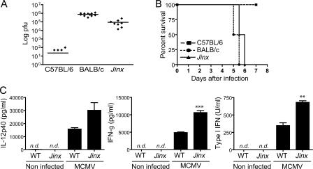
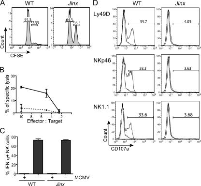
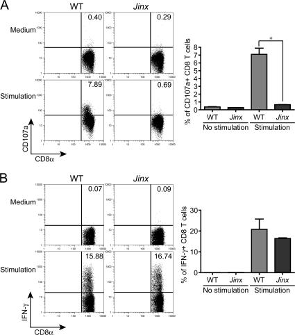
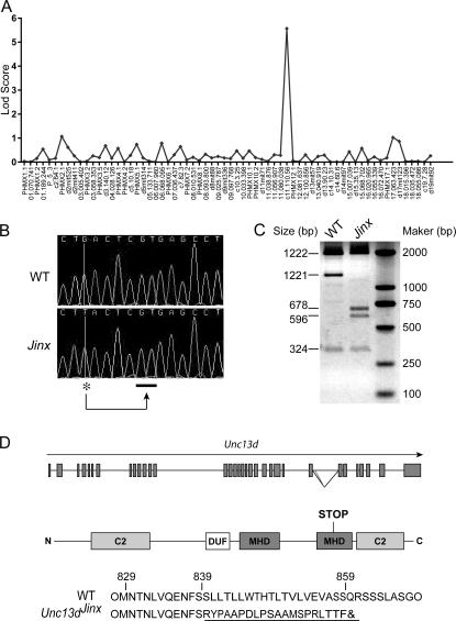
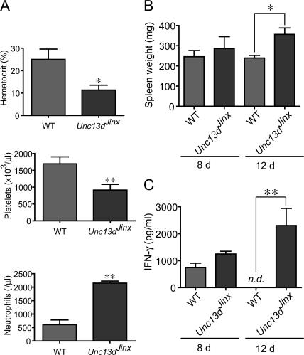
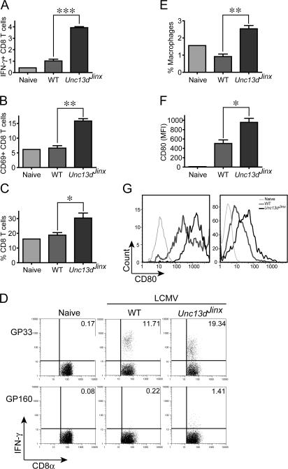
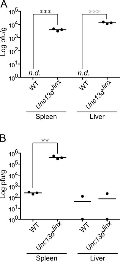
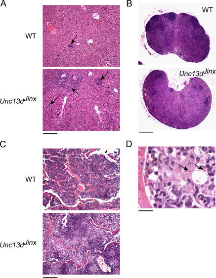
Mouse cytomegalovirus (MCMV) susceptibility often results from defects of natural killer (NK) cell function. Here we describe Jinx, an N-ethyl-N-nitrosourea-induced MCMV susceptibility mutation that permits unchecked proliferation of the virus, causing death. In Jinx homozygotes, activated NK cells and cytotoxic T lymphocytes (CTLs) fail to degranulate, although they retain the ability to produce cytokines, and cytokine levels are markedly elevated in the blood of infected mutant mice. Jinx was mapped to mouse chromosome 11 on a total of 246 meioses and confined to a 4.60-million basepair critical region encompassing 122 annotated genes. The phenotype was ascribed to the creation of a novel donor splice site in Unc13d, the mouse orthologue of human MUNC13-4, in which mutations cause type 3 familial hemophagocytic lymphohistiocytosis (FHL3), a fatal disease marked by massive hepatosplenomegaly, anemia, and thrombocytopenia. Jinx mice do not spontaneously develop clinical features of hemophagocytic lymphohistiocytosis (HLH), but do so when infected with lymphocytic choriomeningitis virus, exhibiting hyperactivation of CTLs and antigen-presenting cells, and inadequate restriction of viral proliferation. In contrast, neither Listeria monocytogenes nor MCMV induces the syndrome. In mice, the HLH phenotype is conditional, which suggests the existence of a specific infectious trigger of FHL3 in humans.


Figures

Figure 1.
Figure 1.
Jinx mutants show high susceptibility and an increase in cytokine production after MCMV infection. (A) PFU were measured in spleens from C57BL/6, BALB/c, and Jinx/Jinx mice on day 5 after the inoculation with 105 PFU of MCMV. BALB/c mice were used as controls for susceptibility. Each point represents an individual animal, and lines refer to means. (B) Time-dependent death of C57BL/6 mice, BALB/c mice, and Jinx/Jinx mutants when challenged with 2.5 × 10⁵ PFU of MCMV. For each genotype, n = 6. The experiment was concluded after 7 d, but no additional deaths were observed for at least 10 additional days. (C) IL-12p40, IFN-γ, and IFN-α/β levels in serum measured 36 h after MCMV infection. n.d., not detected.
Figure 2.
Figure 2.
Jinx NK cells produce IFN-γ after MCMV infection but fail to kill target cells due to a defect in degranulation. (A) In vivo assay of NK cell killing. WT, cells were injected into C57BL/6 mouse; Jinx, cells injected into a Jinx/Jinx homozygote. Numbers refer to the percentage of cells in each gate. (B) Killing of YAC-1 cells in vitro by NK cells purified from MCMV-infected Jinx homozygotes (dashed line) or C57BL/6 cells (solid line). (C) IFN-γ production by NK cells obtained from MCMV-infected C57BL/6 (WT) and Jinx/Jinx homozygotes 2 d after inoculation. Data show the percentage of IFN-γ+ cells among the gated NK1.1+ CD3ɛ population. (D) Stimulus-induced degranulation of NK cells measured by CD107a surface expression. Plate-bound antibodies specific for some NK cell receptors (solid line) or their respective isotype controls (faded line) were used for induction. Numbers indicate the percentage of NK1.1+ CD3ɛ cells expressing CD107a at their surface.
Figure 3.
Figure 3.
Jinx CD8+ T cells produce a normal amount of IFN-γ upon polyclonal stimulation with PMA/ionomycin but fail to degranulate. (A) Surface expression of CD107a. Inset numbers indicate percentage of cells with induced expression. (B) Up-regulation of intracellular IFN-γ. Graphs beneath each FACS illustration show data for three mice.
Figure 4.
Figure 4.
Mapping and positional cloning of Jinx (A) Genome-wide confinement of the phenotype on 30 meioses using a panel of 68 informative markers (bottom). Strongest linkage observed with a marker on chromosome 11, 110.56 Mb. (B) Genomic sequence from a part of intron 26 of Unc13d reveals a G→T transversion (*), which causes splicing to occur distally (underscore), causing incorporation of 53 bp of intronic sequence into exon 26. (C) Effect of the Jinx mutation at the mRNA level. KpnI cuts the WT cDNA twice with a 3,467-bp amplification fragment leading to 1,922-, 1,221-, and 324-bp bands. In Jinx, the 53-bp insertion contains an additional KpnI site, giving 1,922-, 596-, 678-, and 324-bp bands. In the mutant cDNA pool, no WT transcript is detectable. (D) Location of Jinx mutation in the genomic sequence of Unc13d and the structure of the truncated protein predicted from the Jinx mutation. C2, Ca2+-binding domain; MHD, MUNC homology domain; DUF, DUF1041 domain.
Figure 5.
Figure 5.
Unc13dJinx/Jinx mice develop an FHL-like phenotype when infected with LCMV. (A) Hematocrit, platelet count, and neutrophil count in the blood of LCMV-infected WT and Unc13dJinx/Jinx mice 12 d after infection. (B) IFN-γ production in the serum of LCMV-infected WT and Unc13dJinx/Jinx mice 8 and 12 d after infection. n.d., not detected. (C) Spleen weight in WT and Unc13dJinx/Jinx mice 12 d after infection. n = 3 mice per group.
Figure 6.
Figure 6.
Phenotype of CD8+ T cells and APCs in Unc13dJinx/Jinx mutants after LCMV infection. (A–D) Splenocytes from Unc13dJinx/Jinx or WT mice infected with LCMV 12 d previously or uninfected controls were stained for CD8α, CD3ɛ, CD69, and IFN-γ. (A) The percentage of IFN-γ+ CD8+ T cells. (B) Percentage of CD69+ CD8α+ T cells. (C) The percentage of CD8α+ CD3ɛ+ T cells among all splenocytes. (D) Splenocytes were stimulated for 4 h in the presence of LCMV-specific peptide GP33 or the irrelevant peptide GP160. Inset numbers represent the percentage of CD8α+ CD3ɛ+ IFN-γ+ cells. (E) The percentage of macrophages (F4/80+ CD11b+ cells) among all splenocytes. (F) The expression of CD80 (mean fluorescence intensity) by splenic macrophages (gated on the F4/80+ and CD11b+ population). (G) CD80 up-regulation in macrophages (left) and dendritic cells (right) in uninfected mice and in infected WT and Unc13dJinx/Jinx mice. A single uninfected (naive) mouse was used in all experiments, and a representative set of individual mice were used in D and G. For all other panels, n = 3.
Figure 7.
Figure 7.
LCMV titre in the liver and spleen after infection in WT and Unc13dJinx/Jinx mice. Standard plaque assays were performed on spleens and livers 8 (A) and 12 d (B) after LCMV infection. n.d., not detected. n = 3 mice for each group.
Figure 8.
Figure 8.
Histologic appearance of the liver and spleen in LCMV-infected Unc13dJinx/Jinx and WT mice displays features of FHL in different organs. Hematoxylin and eosin staining of sections of livers from LCMV-infected Unc13dJinx/Jinx mice (A) showed an increased number of granulomas of larger size (arrow) compared with livers from infected controls (bar, 200 μm). (B) Infiltration of macrophages is observed in lymph nodes in Unc13dJinx/Jinx mice (arrow), along with a paucity of germinal centers (bar, 0.4 mm). (C) Bone marrow from LCMV-infected Unc13dJinx/Jinx mice is heavily infiltrated by macrophages compared with LCMV-infected WT marrow (bar, 200 μm), and at high magnification (D) RBCs are seen to reside within vesicles in bone marrow macrophages (arrows, hemophagocytosis). Bar, 20 μm.

References

    1. Arase, H., E.S. Mocarski, A.E. Campbell, A.B. Hill, and L.L. Lanier. 2002. Direct recognition of cytomegalovirus by activating and inhibitory NK cell receptors. Science. 296:1323–1326. - PubMed
    1. Lee, S.H., S. Girard, D. Macina, M. Busa, A. Zafer, A. Belouchi, P. Gros, and S.M. Vidal. 2001. Susceptibility to mouse cytomegalovirus is associated with deletion of an activating natural killer cell receptor of the C-type lectin superfamily. Nat. Genet. 28:42–45. - PubMed
    1. Lee, S.H., A. Zafer, Y. de Repentigny, R. Kothary, M.L. Tremblay, P. Gros, P. Duplay, J.R. Webb, and S.M. Vidal. 2003. Transgenic expression of the activating natural killer receptor Ly49H confers resistance to cytomegalovirus in genetically susceptible mice. J. Exp. Med. 197:515–526. - PMC - PubMed
    1. Brown, M.G., A.O. Dokun, J.W. Heusel, H.R. Smith, D.L. Beckman, E.A. Blattenberger, C.E. Dubbelde, L.R. Stone, A.A. Scalzo, and W.M. Yokoyama. 2001. Vital involvement of a natural killer cell activation receptor in resistance to viral infection. Science. 292:934–937. - PubMed
    1. Crozat, K., P. Georgel, S. Rutschmann, N. Mann, X. Du, K. Hoebe, and B. Beutler. 2006. Analysis of the MCMV resistome by ENU mutagenesis. Mamm. Genome. 17:398–406. - PubMed

Publication types

MeSH terms