. 2007 Jun;27(12):4273-82.
doi: 10.1128/MCB.02286-06. Epub 2007 Apr 9.

CDK4 and CDK6 delay senescence by kinase-dependent and p16INK4a-independent mechanisms

Affiliations

CDK4 and CDK6 delay senescence by kinase-dependent and p16INK4a-independent mechanisms

Margarida Ruas et al. Mol Cell Biol. 2007 Jun.

Abstract

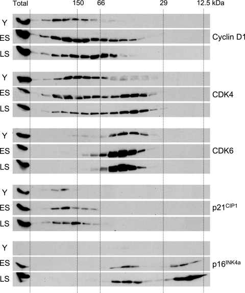
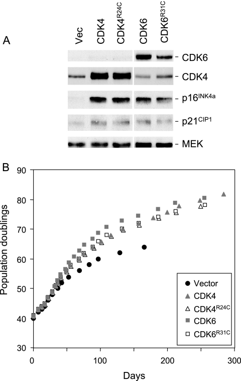
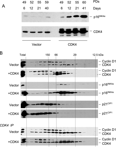
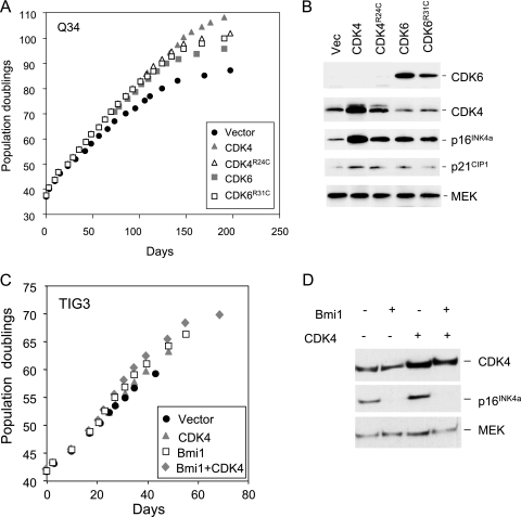
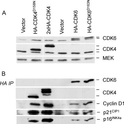
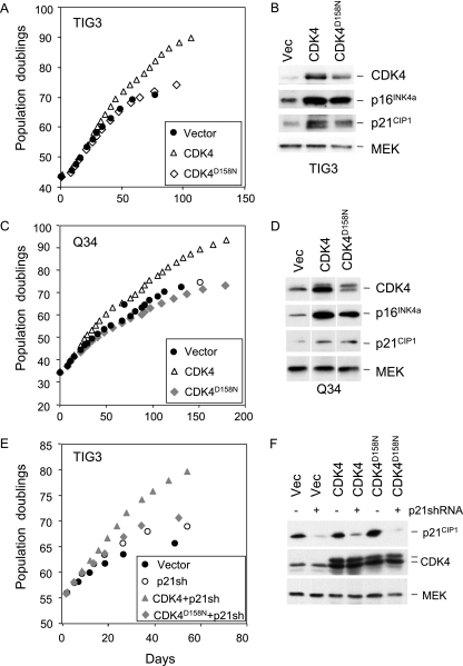
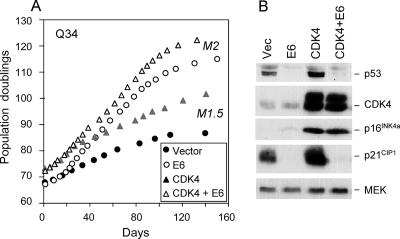
Replicative senescence of human diploid fibroblasts (HDFs) is largely implemented by the cyclin-dependent kinase (CDK) inhibitors p16(INK4a) and p21(CIP1). Their accumulation results in a loss of CDK2 activity, and cells arrest with the retinoblastoma protein (pRb) in its hypophosphorylated state. It has become standard practice to bypass the effects of p16(INK4a) by overexpressing CDK4 or a variant form that is unable to bind to INK4 proteins. Although CDK4 and CDK6 and their INK4-insensitive variants can extend the life span of HDFs, they also cause a substantial increase in the levels of endogenous p16(INK4a). Here we show that CDK4 and CDK6 can extend the life span of HDFs that have inactivating mutations in both alleles of INK4a or in which INK4a levels are repressed, indicating that overexpression of CDK4/6 is not equivalent to ablation of p16(INK4a). However, catalytically inactive versions of these kinases are unable to extend the replicative life span, suggesting that the impact of ectopic CDK4/6 depends on their ability to phosphorylate as yet unidentified substrates rather than to sequester CDK inhibitors. Since p16(INK4a) deficiency, CDK4 expression, and p53 or p21(CIP1) ablation have additive effects on replicative life span, our results underscore the idea that senescence is an integrated response to diverse signals.


Figures

FIG. 1.
FIG. 1.
Comparison of cyclin D1-CDK-CKI complexes in young and senescent HDFs. Equal amounts of cell lysate (2.5 mg) from young (Y), early senescence (ES), or late senescence (LS) TIG3 cells were subjected to gel filtration chromatography on a Superdex 200 column. Equivalent amounts (1/20th) of the individual fractions were separated by SDS-PAGE in a 12% acrylamide gel and immunoblotted with antisera against cyclin D1, CDK4, CDK6, p21CIP1, and p16INK4a. The lane on the left of each panel corresponds to a sample (12.5 μg) of total protein analyzed directly (i.e., without gel filtration).
FIG. 2.
FIG. 2.
Extension of HDF life span by wild-type and mutant versions of CDK4 and CDK6. (A) Hs68 cells were infected at PD40 with retroviruses encoding wild-type CDK4, the R24C mutant of CDK4, wild-type CDK6, the R31C mutant of CDK6, or the empty vector as indicated. Following selection in puromycin, the cell pools were analyzed by immunoblotting with antibodies against CDK4, CDK6, p16INK4a, and p21CIP1. MEK was used as a control for loading. Note that the samples were analyzed on the same gel, but the scanned images were subsequently edited for continuity of presentation. (B) Infected cell pools were passaged under standard conditions of tissue culture until they reached M1 senescence, as judged by failure to double in 4 weeks. The curves show cumulative PDs at each time point.
FIG. 3.
FIG. 3.
Induction of p16INK4a by ectopic CDK4 and its effect on cyclin D-CDK complexes. (A) TIG3 cells were infected with a retrovirus encoding wild-type CDK4 or the empty-vector control. Following drug selection (6 days), the cells were passaged under standard conditions and cell lysates prepared at various time points. Cumulative PDs at each time point are as indicated. Samples (30 μg) of total protein were fractionated by SDS-PAGE and immunoblotted for p16INK4a and CDK4. (B) Extracts from TIG3 cells infected with a retrovirus encoding wild-type CDK4 or the empty-vector control were subjected to gel filtration and analyzed as in Fig. 1. Cyclin D1 and CDK4 were visualized on the same immunoblot, with the upper band corresponding to cyclin D1. Ectopic expression of CDK4 resulted in two immunoreactive bands, as discussed in the text. In the lower panels, samples of each fraction were immunoprecipitated with an antibody against CDK4 prior to SDS-PAGE and immunoblotting.
FIG. 4.
FIG. 4.
CDK4 and CDK6 can extend the life span of p16INK4a-deficient HDFs. (A) The Q34 strain of p16INK4a-deficient HDFs was infected at PD37 with retroviruses encoding wild-type CDK4, the R24C mutant of CDK4, wild-type CDK6, the R31C mutant of CDK6, or the empty vector, as indicated. Following selection, the infected cell pools were passaged under standard conditions of tissue culture until they reached M1 senescence. Curves show cumulative PDs at each time point. (B) Lysates prepared from infected cell pools were analyzed by immunoblotting with antibodies against CDK4, CDK6, p16INK4a, and p21CIP1. MEK was used as a control for loading. (C and D) The TIG3 strain of HDFs was infected at PD42 with retroviruses encoding CDK4 or Bmi1 or both, along with appropriate vector controls. Following selection, the infected cell pools were passaged under standard conditions of tissue culture until they reached M1 senescence. The curves show cumulative PDs at each time point. Lysates prepared from the infected cell pools were analyzed by immunoblotting with antibodies against CDK4, p16INK4a, and MEK.
FIG. 5.
FIG. 5.
Catalytically inactive versions of CDK4 and CDK6 are able to associate with cyclin D1, p21CIP1, and p16INK4a. TIG3 cells at PD42 were infected with retroviruses encoding HA-tagged versions of wild-type CDK4, CDK4D158N, wild-type CDK6, or CDK6D163N or empty-vector controls. (A) Following drug selection, cell lysates were prepared and samples (25 μg) of total protein were analyzed by SDS-PAGE in a 12% gel and immunoblotted with antibodies against CDK4 and CDK6. MEK served as a control for loading. As discussed in the text, ectopic expression of CDK4 resulted in multiple bands, the exact provenance of which remains unclear. Note also that the wild-type CDK4 construct contained two copies of the HA epitope. (B) Samples (500 μg) of protein were immunoprecipitated with a monoclonal antibody against the HA epitope, fractionated by SDS-PAGE, and immunoblotted for CDK4, CDK6, cyclin D1, p21CIP1, or p16INK4a as indicated.
FIG. 6.
FIG. 6.
Catalytically inactive CDK4 does not extend the life span of HDFs. TIG3 cells (A and B) or Q34 cells (C and D) were infected with retroviruses encoding wild-type CDK4, the kinase-dead version, CDK4D158N, or empty vector. The infected cell pools were passaged under standard conditions of tissue culture until they reached M1 senescence. The curves (A and C) show cumulative PDs at each time point. Samples of cell lysate were immunoblotted with antibodies against CDK4, p16INK4a, and p21CIP1 (B and D). MEK was used as a control for loading. (E and F) TIG3 cells transduced with either CDK4 or CDK4D158N or the empty vector were infected with a retrovirus containing shRNA against p21CIP1. The curves show cumulative PDs at each time point (E). Samples of cell lysate were immunoblotted with antibodies against p21CIP1, CDK4, and MEK (F).
FIG. 7.
FIG. 7.
Effects of p53 ablation and CDK4 overexpression are additive. (A) Q34 cells that had been previously infected with a retrovirus encoding wild-type CDK4 or vector-only control cells were superinfected with a virus encoding the E6 protein from HPV16. No drug selection was required because of the proliferative advantage gained by cells that express E6. The infected cell pools were passaged under standard conditions until cultures failed to double in 4 weeks. The curves show cumulative PDs at each time point. Note that cultures expressing E6 ended with an M2 phenotype, as demonstrated by BrdU incorporation and extensive cell death. The phenotype of the control cultures was indistinguishable from M1. (B) Samples of cell lysate were analyzed by immunoblotting for p53, CDK4, p16INK4a, or p21CIP1, as indicated. MEK was used as a control for loading.

References

    1. Alcorta, D. A., Y. Xiong, D. Phelps, G. Hannon, D. Beach, and J. C. Barrett. 1996. Involvement of the cyclin-dependent kinase inhibitor p16 (INK4a) in replicative senescence of normal human fibroblasts. Proc. Natl. Acad. Sci. USA 93:13742-13747. - PMC - PubMed
    1. Baker, G. L., M. W. Landis, and P. W. Hinds. 2005. Multiple functions of D-type cyclins can antagonize pRb-mediated suppression of proliferation. Cell Cycle 4:330-338. - PubMed
    1. Bates, S., L. Bonetta, D. MacAllan, D. Parry, A. Holder, C. Dickson, and G. Peters. 1994. CDK6 (PLSTIRE) and CDK4 (PSK-J3) are a distinct subset of the cyclin-dependent kinases that associate with cyclin D1. Oncogene 9:71-79. - PubMed
    1. Berthet, C., E. Aleem, V. Coppola, L. Tessarollo, and P. Kaldis. 2003. Cdk2 knockout mice are viable. Curr. Biol. 13:1775-1785. - PubMed
    1. Bond, J. A., M. F. Haughton, J. M. Rowson, P. J. Smith, V. Gire, D. Wynford-Thomas, and F. S. Wyllie. 1999. Control of replicative life span in human cells: barriers to clonal expansion intermediate between M1 senescence and M2 crisis. Mol. Cell. Biol. 19:3103-3114. - PMC - PubMed

MeSH terms

Substances