. 2007 Apr 9;177(1):115-25.
doi: 10.1083/jcb.200609161.

Grd19/Snx3p functions as a cargo-specific adapter for retromer-dependent endocytic recycling

Affiliations

Grd19/Snx3p functions as a cargo-specific adapter for retromer-dependent endocytic recycling

Todd I Strochlic et al. J Cell Biol. .

Abstract

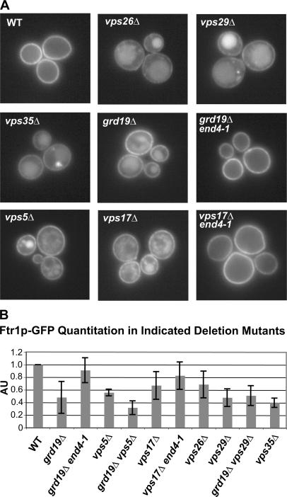
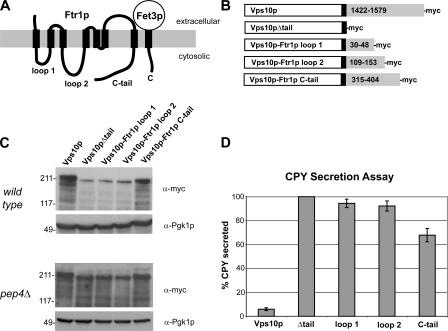
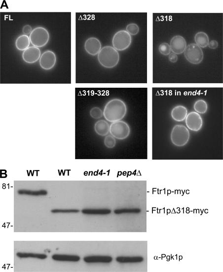
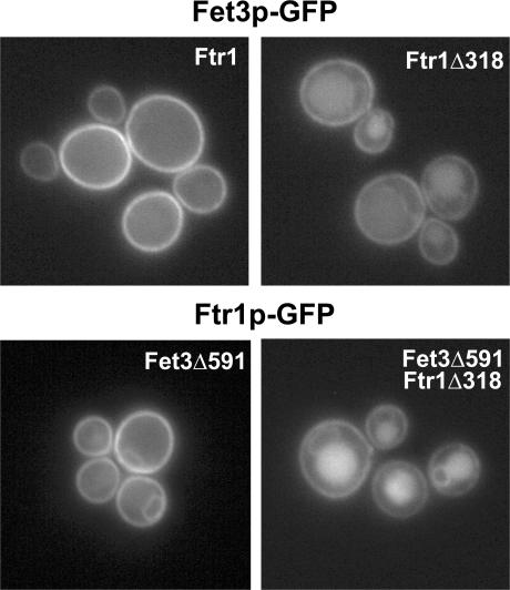
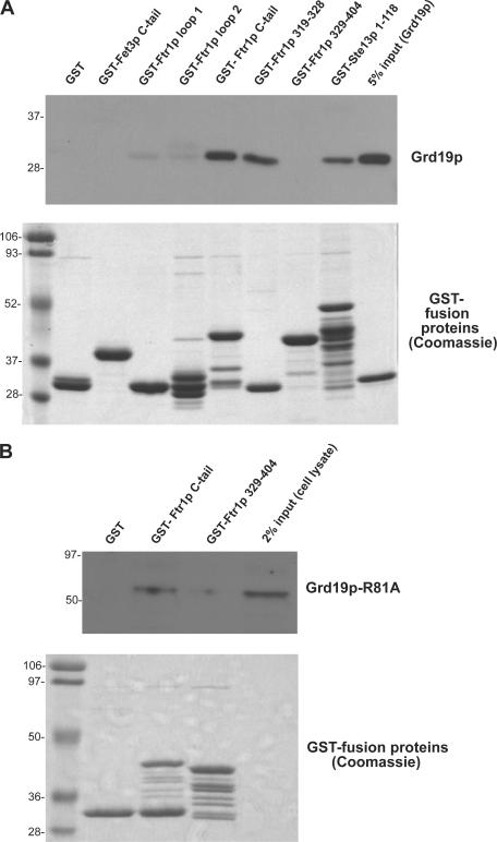
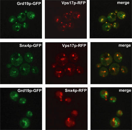
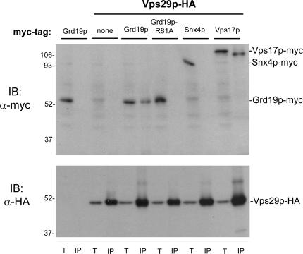
Amajor function of the endocytic system is the sorting of cargo to various organelles. Endocytic sorting of the yeast reductive iron transporter, which is composed of the Fet3 and Ftr1 proteins, is regulated by available iron. When iron is provided to iron-starved cells, Fet3p-Ftr1p is targeted to the lysosome-like vacuole and degraded. In contrast, when iron is not available, Fet3p-Ftr1p is maintained on the plasma membrane via an endocytic recycling pathway requiring the sorting nexin Grd19/Snx3p, the pentameric retromer complex, and the Ypt6p Golgi Rab GTPase module. A recycling signal in Ftr1p was identified and found to bind directly to Grd19/Snx3p. Retromer and Grd19/Snx3p partially colocalize to tubular endosomes, where they are physically associated. After export from the endosome, Fet3p-Ftr1p transits through the Golgi apparatus for resecretion. Thus, Grd19/Snx3p, functions as a cargo-specific adapter for the retromer complex, establishing a precedent for a mechanism by which sorting nexins expand the repertoire of retromer-dependent cargos.


Figures

Figure 1.
Figure 1.
Retromer and the sorting nexin Grd19p are required to maintain PM localization of Fet3p–Ftr1p under iron-limiting conditions. (A) Ftr1p-GFP is mislocalized to the lumen of the vacuole in cells that lack a functional retromer complex (vps5Δ, vps17Δ, vps26Δ, vps29Δ, and vps35Δ) or the sorting nexin Grd19p (grd19Δ). Cells expressing Ftr1p-GFP from the native FTR1 locus were grown in iron-deficient medium and analyzed by fluorescence microscopy. (B) Quantitative immunoblot analysis of steady-state levels of Ftr1p-GFP in wild-type (WT) cells and the indicated deletion mutants. Cells were grown in iron-deficient medium, and whole-cell extracts were prepared and immunoblotted with an anti-GFP antibody and an antibody to Pgk1p (3-phosphoglycerate kinase) which was used as a loading control. ECF was used to detect the antibodies, and the ECF signals were quantified using ImageQuant software and normalized to the loading control. AU, arbitrary units. The amount of Ftr1p-GFP detected from the wild-type strain was set to 1.0, and the means and SDs for each of the indicated strains were calculated from three independent experiments.
Figure 2.
Figure 2.
The C-terminal cytoplasmic tail of Ftr1p contains an endosome-to-Golgi sorting/recycling signal. (A) The predicted topology of the S. cerevisiae high-affinity iron transporter. The iron oxidase Fet3p contains a single transmembrane domain, whereas Ftr1p, the iron permease, contains seven transmembrane domains connected by three cytoplasmic loops. Both proteins have cytoplasmic C-terminal tails. (B) Control proteins and chimeric Vps10p-Ftr1p proteins were generated to identify a potential recycling signal within Ftr1p. The indicated cytoplasmic regions of Ftr1p (numbers in gray rectangles) were fused to Vps10p just downstream of the single transmembrane domain (black rectangle). All constructs also contained a myc-epitope tag at the C terminus. (C) Steady-state immunoblot analysis of myc-tagged Vps10p-Ftr1p chimeras and control constructs. Whole-cell extracts were prepared and immunoblotted with an anti-myc antibody and an antibody to Pgk1p. An identical analysis of the abundance of the chimeric proteins was done with pep4Δ cells, which are deficient in vacuolar proteolysis. (D) Analysis of CPY secretion from Vps10p-Ftr1p strains by quantitative colony blot assay. Secretion of vacuolar CPY from strains expressing chimeric Vps10p-Ftr1p fusion proteins was quantified by colony immunoblotting with an anti-CPY monoclonal antibody. ECF was used to detect anti-CPY antibody, and the ECF signals were quantified using ImageQuant software. The amount of CPY secreted from the Vps10Δtail strain was set to 100%, and the means and SDs for each of the indicated strains were calculated from 10 independent measurements.
Figure 3.
Figure 3.
The Ftr1p recycling signal resides between residues 319–328. (A) Localization of GFP-tagged Ftr1p truncation mutants. Cells were grown in iron-deficient medium and imaged by fluorescence microscopy. Numbers indicate the last Ftr1p residue expressed (top). An internal deletion mutant lacking aa 319–328 was also tested (Δ319–328). (B) Analysis of steady-state levels of full-length Ftr1p-myc and Ftr1pΔ318-myc grown in iron-deficient medium. Whole-cell extracts were prepared from the indicated strains and were immunoblotted with an anti-myc antibody and an antibody to Pgk1p as a loading control.
Figure 4.
Figure 4.
The Ftr1p recycling signal is required for endocytic recycling of Fet3p. Yeast strains were constructed in which Fet3p-GFP was expressed in cells expressing full-length Ftr1p or Ftr1pΔ318 (top), or Ftr1p-GFP and Ftr1pΔ318-GFP in cells expressing a C-tail–truncated form of Fet3p, Fet3pΔ591 (bottom). The cells were grown in iron-deficient medium and imaged by fluorescence microscopy.
Figure 5.
Figure 5.
Grd19p binds directly to the Ftr1p recycling signal. (A) The following GST-fusion proteins were expressed and purified from bacteria and immobilized on glutathione–Sepharose beads: GST-Fet3p cytoplasmic tail (residues 581–636), GST-Ftr1p cytoplasmic loop 1 (residues 30–48), GST-Ftr1p cytoplasmic loop 2 (residues 109–153), GST-Ftr1p cytoplasmic tail (residues 315–404), GST-Ftr1p(319–328), which is the putative recycling signal, and GST-Ftr1p(329–404), which contains the entire C-terminal domain except for the recycling signal. Purified fusion proteins were incubated with purified recombinant His6-Grd19p, which contains a T7 epitope tag on the N terminus. The beads were washed, and bound proteins were eluted in SDS sample buffer. After SDS-PAGE, Grd19p was detected using an anti-T7 epitope antibody (top) and the GST fusion proteins were visualized by Coomassie blue staining (bottom). (B) Yeast strains expressing Grd19p-R81A-myc and the indicated GST fusion proteins were grown in synthetic media containing 4% galactose. Cells were harvested, converted to spheroplasts, and the bifunctional chemical cross-linking reagent DSP was added to a final concentration of 4 mM. The cells were then lysed, and the GST fusion proteins were captured on glutathione–Sepharose beads. The cross-links were disrupted by reduction in SDS sample buffer, and the samples were probed by immunoblotting with anti-myc antibodies (top). GST fusion proteins were visualized by Coomassie blue staining (bottom).
Figure 6.
Figure 6.
Retromer and Grd19p partially colocalize on tubular endosomal membranes. Live cells coexpressing endogenous C-terminally tagged Grd19p-GFP and Vps17p-RFP, Snx4p-GFP and Vps17p-RFP, and Grd19p-GFP and Snx4p-RFP were imaged by spinning disc confocal microscopy. Images acquired using the individual red and green channels were then merged to determine extent of colocalization. Shown are two-dimensional maximum projections of the entire cell volumes.
Figure 7.
Figure 7.
Grd19p copurifies with retromer after chemical cross-linking. Strains expressing the indicated epitope-tagged proteins were grown in synthetic medium and converted to spheroplasts, and DSP was added to a final concentration of 5 mM. The cells were lysed, and Vps29-HA was captured on anti-HA beads. The cross-links were disrupted by reduction in SDS sample buffer, and the samples were probed by immunoblotting with anti-myc (top) or anti-HA antibodies (bottom). Note that the 9E10 (anti-myc) antibody detects a faint 55-kD background band that migrates just above Grd19p-myc, and that Vps17p-myc recovered by immunoprecipitation runs ∼10 kD below Vps17p-myc detected in whole-cell extract. T, 2% total (whole-cell extract); IP, immunoprecipitates.

Similar articles

Cited by

References

    1. Arighi, C.N., L.M. Hartnell, R.C. Aguilar, C.R. Haft, and J.S. Bonifacino. 2004. Role of the mammalian retromer in sorting of the cation-independent mannose 6-phosphate receptor. J. Cell Biol. 165:123–133. - PMC - PubMed
    1. Askwith, C., D. Eide, A. Van Ho, P.S. Bernard, L. Li, S. Davis-Kaplan, D.M. Sipe, and J. Kaplan. 1994. The FET3 gene of S. cerevisiae encodes a multicopper oxidase required for ferrous iron uptake. Cell. 76:403–410. - PubMed
    1. Bensen, E.S., B.G. Yeung, and G.S. Payne. 2001. Ric1p and the Ypt6p GTPase function in a common pathway required for localization of trans-Golgi network membrane proteins. Mol. Biol. Cell. 12:13–26. - PMC - PubMed
    1. Blondel, M.O., J. Morvan, S. Dupre, D. Urban-Grimal, R. Haguenauer-Tsapis, and C. Volland. 2004. Direct sorting of the yeast uracil permease to the endosomal system is controlled by uracil binding and Rsp5p-dependent ubiquitylation. Mol. Biol. Cell. 15:883–895. - PMC - PubMed
    1. Bonifacino, J.S., and R. Rojas. 2006. Retrograde transport from endosomes to the trans-Golgi network. Nat. Rev. Mol. Cell Biol. 7:568–579. - PubMed

Publication types

MeSH terms