

Deficiency in the LIM-only protein Fhl2 impairs skin wound healing
- PMID: 17420295
- PMCID: PMC2064120
- DOI: 10.1083/jcb.200606043
Deficiency in the LIM-only protein Fhl2 impairs skin wound healing
Abstract
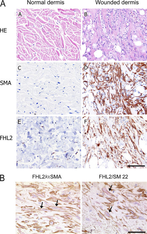
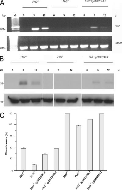
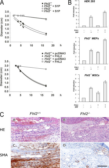
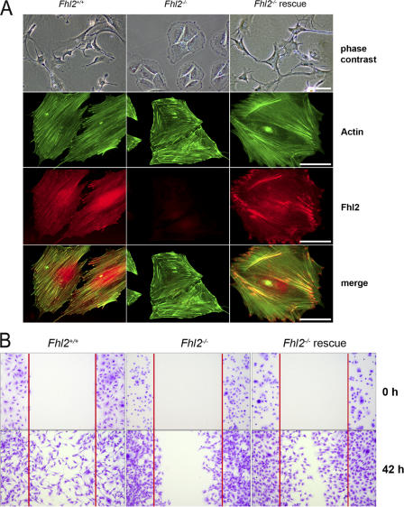
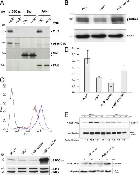
After skin wounding, the repair process is initiated by the release of growth factors, cytokines, and bioactive lipids from injured vessels and coagulated platelets. These signal molecules induce synthesis and deposition of a provisional extracellular matrix, as well as fibroblast invasion into and contraction of the wounded area. We previously showed that sphingosine-1-phosphate (S1P) triggers a signal transduction cascade mediating nuclear translocation of the LIM-only protein Fhl2 in response to activation of the RhoA GTPase (Muller, J.M., U. Isele, E. Metzger, A. Rempel, M. Moser, A. Pscherer, T. Breyer, C. Holubarsch, R. Buettner, and R. Schule. 2000. EMBO J. 19:359-369; Muller, J.M., E. Metzger, H. Greschik, A.K. Bosserhoff, L. Mercep, R. Buettner, and R. Schule. 2002. EMBO J. 21:736-748.). We demonstrate impaired cutaneous wound healing in Fhl2-deficient mice rescued by transgenic expression of Fhl2. Furthermore, collagen contraction and cell migration are severely impaired in Fhl2-deficient cells. Consequently, we show that the expression of alpha-smooth muscle actin, which is regulated by Fhl2, is reduced and delayed in wounds of Fhl2-deficient mice and that the expression of p130Cas, which is essential for cell migration, is reduced in Fhl2-deficient cells. In summary, our data demonstrate a function of Fhl2 as a lipid-triggered signaling molecule in mesenchymal cells regulating their migration and contraction during cutaneous wound healing.
Figures
Similar articles
-
Impaired intestinal wound healing in Fhl2-deficient mice is due to disturbed collagen metabolism.Exp Cell Res. 2008 Dec 10;314(20):3684-91. doi: 10.1016/j.yexcr.2008.09.023. Epub 2008 Oct 10. Exp Cell Res. 2008. PMID: 18950620
-
Four-and-a-half LIM domain protein 2 is a novel regulator of sphingosine 1-phosphate receptor 1 in CCL19-induced dendritic cell migration.J Immunol. 2010 Aug 1;185(3):1466-75. doi: 10.4049/jimmunol.0903449. Epub 2010 Jun 30. J Immunol. 2010. PMID: 20592280
-
Deficiency in the LIM-only protein FHL2 impairs assembly of extracellular matrix proteins.FASEB J. 2008 Jul;22(7):2508-20. doi: 10.1096/fj.07-095521. Epub 2008 Mar 20. FASEB J. 2008. PMID: 18356303
-
The biological relevance of FHL2 in tumour cells and its role as a putative cancer target.Anticancer Res. 2007 Jan-Feb;27(1A):55-61. Anticancer Res. 2007. PMID: 17352216 Review.
-
The role of FHL2 in wound healing and inflammation.FASEB J. 2019 Jul;33(7):7799-7809. doi: 10.1096/fj.201802765RR. Epub 2019 Apr 2. FASEB J. 2019. PMID: 30939249 Review.
Cited by
-
Deficiency in four and one half LIM domain protein 2 (FHL2) aggravates liver fibrosis in mice.BMC Gastroenterol. 2013 Jan 14;13:8. doi: 10.1186/1471-230X-13-8. BMC Gastroenterol. 2013. PMID: 23311569 Free PMC article.
-
The LIM-only protein FHL2 mediates ras-induced transformation through cyclin D1 and p53 pathways.PLoS One. 2008;3(11):e3761. doi: 10.1371/journal.pone.0003761. Epub 2008 Nov 19. PLoS One. 2008. PMID: 19018287 Free PMC article.
-
Protective transcriptional mechanisms in cardiomyocytes and cardiac fibroblasts.J Mol Cell Cardiol. 2019 Jul;132:1-12. doi: 10.1016/j.yjmcc.2019.04.023. Epub 2019 Apr 28. J Mol Cell Cardiol. 2019. PMID: 31042488 Free PMC article. Review.
-
FHL2 expression by cancer-associated fibroblasts promotes metastasis and angiogenesis in lung adenocarcinoma.Int J Cancer. 2025 Jan 15;156(2):431-446. doi: 10.1002/ijc.35174. Epub 2024 Sep 8. Int J Cancer. 2025. PMID: 39244734 Free PMC article.
-
Mammalian Actin-binding Protein-1/Hip-55 Interacts with FHL2 and Negatively Regulates Cell Invasion.J Biol Chem. 2016 Jul 1;291(27):13987-13998. doi: 10.1074/jbc.M116.725739. Epub 2016 Apr 26. J Biol Chem. 2016. PMID: 27129278 Free PMC article.
References
-
- Ashcroft, G.S., X. Yang, A.B. Glick, M. Weinstein, J.L. Letterio, D.E. Mizel, M. Anzano, T. Greenwell-Wild, S.M. Wahl, C. Deng, and A.B. Roberts. 1999. Mice lacking Smad3 show accelerated wound healing and an impaired local inflammatory response. Nat. Cell Biol. 1:260–266. - PubMed
-
- Chai, J., and A.S. Tarnawski. 2002. Serum response factor: discovery, biochemistry, biological roles and implications for tissue injury healing. J. Physiol. Pharmacol. 53:147–157. - PubMed
-
- Friedrichs, N., R. Jager, E. Paggen, C. Rudlowski, S. Merkelbach-Bruse, H. Schorle, and R. Buettner. 2005. Distinct spatial expression patterns of AP-2alpha and AP-2gamma in non-neoplastic human breast and breast cancer. Mod. Pathol. 18:431–438. - PubMed
Publication types
MeSH terms
Substances
LinkOut - more resources
Full Text Sources
Molecular Biology Databases
