. 2007 Apr 17;104(16):6782-7.
doi: 10.1073/pnas.0701999104. Epub 2007 Apr 9.

Phosphorylation-dependent regulation of nuclear localization and functions of integrin-linked kinase

Affiliations

Phosphorylation-dependent regulation of nuclear localization and functions of integrin-linked kinase

Filippo Acconcia et al. Proc Natl Acad Sci U S A. .

Abstract

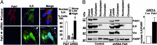
Integrin-linked kinase (ILK) is a phosphorylated protein that regulates physiological processes that overlap with those regulated by p21-activated kinase 1 (PAK1). Here we report the possible role of ILK phosphorylation by PAK1 in ILK-mediated signaling and intracellular translocation. We found that PAK1 phosphorylates ILK at threonine-173 and serine-246 in vitro and in vivo. Depletion of PAK1 decreased the levels of endogenous ILK phosphorylation in vivo. Mutation of PAK1 phosphorylation sites on ILK to alanine reduced cell motility and cell proliferation. Biochemical fractionation, confocal microscopy, and chromatin-interaction analyses of human cells revealed that ILK localizes predominantly in the cytoplasm but also resides in the nucleus. Transfection of MCF-7 cells with point mutants ILK-T173A, ILK-S246A, or ILK-T173A; S246A (ILK-DM) altered ILK localization. Selective depletion of PAK1 dramatically increased the nuclear and focal point accumulation of ILK, further demonstrating a role for PAK1 in ILK translocation. We also identified functional nuclear localization sequence and nuclear export sequence motifs in ILK, delineated an apparently integral role for ILK in maintaining normal nuclear integrity, and established that ILK interacts with the regulatory region of the CNKSR3 gene chromatin to negatively modulate its expression. Together, these results suggest that ILK is a PAK1 substrate, undergoes phosphorylation-dependent shuttling between the cell nucleus and cytoplasm, and interacts with gene-regulatory chromatin.


Conflict of interest statement

The authors declare no conflict of interest.

Figures

Fig. 1.
Fig. 1.
ILK phosphorylation. (A) Potential PAK1 phosphorylation sites. (B and C) In vitro PAK1 phosphorylation of ILK (B) and ILK point mutant (C) constructs. Ponceau S staining indicates equal loading. (D) In vivo phosphorylation of ILK-WT and ILK-DM proteins in the respective MCF-7 clones. (E) Repression of PAK1 expression levels in MCF-7 cells stably expressing PAK1 shRNA. (F) Loss of in vivo phosphorylation of ILK in MCF-7 cells on depletion of PAK1 expression levels by stably overexpressing PAK1-specific shRNA. ILK bands are indicated by asterisks.
Fig. 2.
Fig. 2.
Role of PAK1 phosphorylation sites on ILK cellular activities and localization. (A and B) Cell growth curve (A) and Boyden chamber assays (B) in exponentially growing (10% serum) MCF-7/pcDNA, ILK-WT, and ILK-DM clones. Data are the means ± SD of three independent experiments. ∗, differences from MCF-7/pcDNA cells; #, differences from ILK-WT #21 cells. Analyses included using the Student t test for overall significant differences within groups (P < 0.05). (C) MCF-7 cells transiently transfected with ILK-expressing vectors as labeled were fixed and stained for V5 tag. (Scale bars: 20 μm.) (D) Western blot analysis of exponentially growing Hec1A and MCF-7 cells after subcellular fractionation. Blots show representative results of three different experiments. ∗ indicates ILK in the nuclear fraction.
Fig. 3.
Fig. 3.
ILK NLS and NES. MCF-7 cells transiently transfected with the V5-ILK NLS mutant (K363A) (A) or the V5-ILK NES mutant (I400A) (B) expressing vectors as labeled were fixed and stained for V5 tag (red), the F-actin-binding protein phalloidin (green), and DNA (blue). (Scale bars: 20 μm.) Transfected cells frequently displayed abnormalities (arrows).
Fig. 4.
Fig. 4.
Role of PAK1 in ILK nuclear localization. (A) MCF-7 cells were transfected with either scrambled oligonucleotides or PAK1 siRNA. After 48 h, cells were fixed and stained for endogenous ILK (green), PAK1 (red), and DNA (blue). (Scale bars: 20 μm.) (B) Western blot analysis of exponentially growing control vector and PAK1 shRNA stably transfected cells that were subjected to subcellular fractionation. Vin, vinculin. Blots show representative results of two different experiments. The graph shows densitometric analyses.
Fig. 5.
Fig. 5.
Role of PAK1 in ILK nuclear export. (A) MCF-7 cells were serum-starved for 48 h and treated with EGF (100 ng/ml) or leptomycin B (2 ng/ml) for 1 h, and lysates from different subcellular fractions were immunoblotted with the indicated antibodies. (B) Immunofluorescent staining of MCF-7 cells. Cells were treated as described above and immunostained for the endogenous ILK (green), F-actin-binding protein phalloidin (red), and DNA (blue). (Scale bars: 20 μm.) The results shown in B are quantified in SI Table 1.
Fig. 6.
Fig. 6.
ILK nuclear functions. (A) pcDNA/MCF-7, ILK-WT #21, and ILK-DM #7 clones were fixed and stained for lamin A/C and DNA. The results represented here are quantified in SI Table 2. Nuclear abnormalities were scored in at least 200 cells. (B) Schematic representation of the ILK-chromatin target region for the CNKSR3 gene. (C Upper) ChIP with V5 antibody validated the association of ILK-WT and ILK-DM with the CKNSR3 regulatory chromatin. (C Lower) ChIP with ILK antibody shows similar chromatin association of ILK in HEC1A cells. (D) Association of ILK-WT and ILK-DM with chromatin region 500 bp proximal to the CNKSR3 transcription start site. (E) Luciferase assay in MCF-7 cells shows the possible transcription-repressing functions of the ILK target chromatin. (F and G) Repressive effects of ILK-WT and ILK-DM overexpression on PGL3-ILK chromatin luciferase in the respective MCF-7 clones (H). Silencing of ILK expression in MCF-7 cell results in the relieving of repression on PGL3-ILK chromatin luciferase. Western blotting shows efficient silencing of ILK by siRNA (H Inset). (I) Luciferase assay in MCF-7 cells shows the effect of ILK-WT, ILK-DM, and ILK-NLS mutant proteins on PGL3-ILK target chromatin luciferase. (J) RT-PCR analysis of CNKSR3 expression in MCF-7 pcDNA, ILK-WT, and ILK-DM clones shows distinct repression of the gene by ILK. Quantitation from two different experiments is provided below. Data are the means ± SD of three independent experiments. ∗, Significantly different from values for the respective controls (P < 0.05, Student's t test for differences within groups).

References

    1. Hannigan G, Troussard AA, Dedhar S. Nat Rev Cancer. 2005;5:51–63. - PubMed
    1. Esfandiarei M, Suarez A, Amaral A, Si X, Rahmani M, Dedhar S. Circ Res. 2006;99:354–361. - PubMed
    1. Wang B, Yurecko RS, Dedhar S, Cleary PP. Cell Microbiol. 2006;8:257–266. - PubMed
    1. Legate KR, Montanez E, Kudlacek O, Fassler R. Nat Rev Mol Cell Biol. 2006;7:20–31. - PubMed
    1. Driver GA, Veale RB. Cancer Cell Int. 2006;27:6–12. - PMC - PubMed

Publication types

MeSH terms